Institut für Molekularbiologie, OE5250, Medizinische Hochschule Hannover, Hannover, Germany.
PLoS One. 2012;7(9):e45100. doi: 10.1371/journal.pone.0045100. Epub 2012 Sep 11.
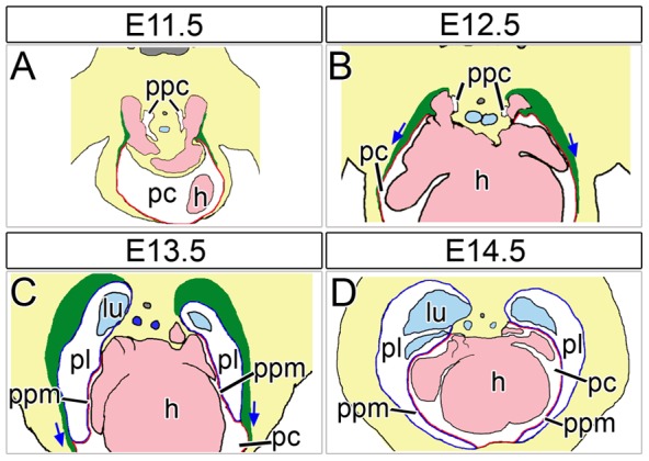
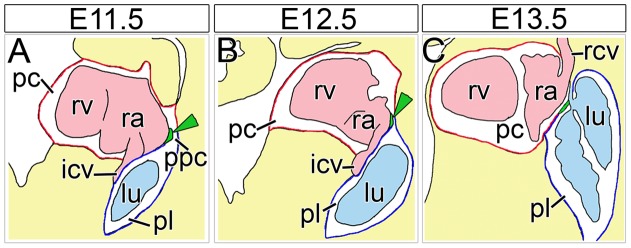
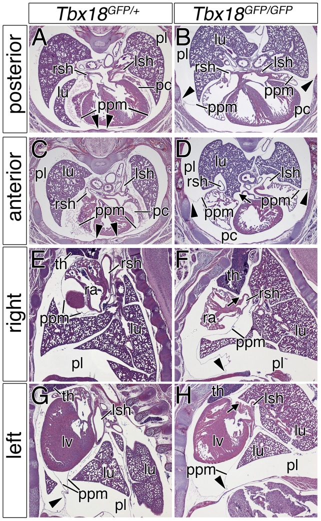
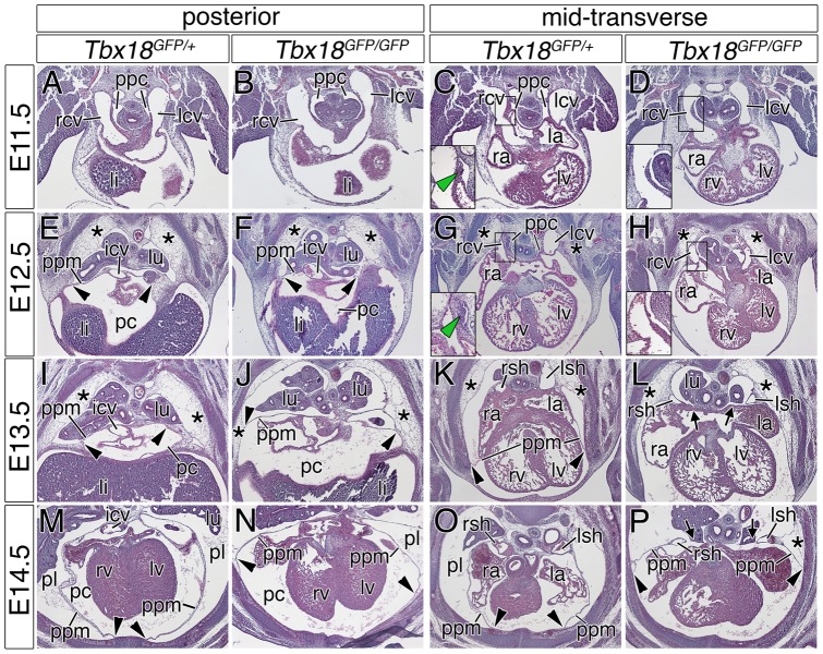
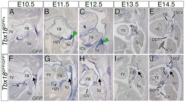
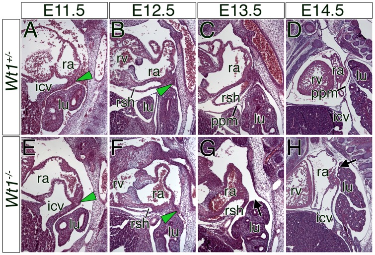
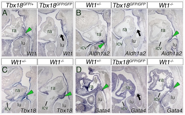
The pleuropericardial membranes are fibro-serous walls that separate the pericardial and pleural cavities and anchor the heart inside the mediastinum. Partial or complete absence of pleuropericardial membranes is a rare human disease, the etiology of which is poorly understood. As an attempt to better understand these defects, we wished to analyze the cellular and molecular mechanisms directing the separation of pericardial and pleural cavities by pleuropericardial membranes in the mouse. We found by histological analyses that both in Tbx18- and Wt1-deficient mice the pleural and pericardial cavities communicate due to a partial absence of the pleuropericardial membranes in the hilus region. We trace these defects to a persisting embryonic connection between these cavities, the pericardioperitoneal canals. Furthermore, we identify mesenchymal ridges in the sinus venosus region that tether the growing pleuropericardial membranes to the hilus of the lung, and thus, close the pericardioperitoneal canals. In Tbx18-deficient embryos these mesenchymal ridges are not established, whereas in Wt1-deficient embryos the final fusion process between these tissues and the body wall does not occur. We suggest that this fusion is an active rather than a passive process, and discuss the interrelation between closure of the pericardioperitoneal canals, lateral release of the pleuropericardial membranes from the lateral body wall, and sinus horn development.
胸膜心包膜是分隔心包腔和胸膜腔并将心脏固定在纵隔内的纤维浆膜壁。胸膜心包膜部分或完全缺失是一种罕见的人类疾病,其病因尚不清楚。为了更好地理解这些缺陷,我们试图分析细胞和分子机制,指导小鼠胸膜心包膜将心包腔和胸膜腔分隔开。通过组织学分析,我们发现 Tbx18 和 Wt1 缺陷型小鼠的胸膜腔和心包腔由于在肺门区域胸膜心包膜部分缺失而相通。我们将这些缺陷追溯到这些腔室(心包腹膜管)之间持续存在的胚胎连接。此外,我们在窦静脉区域发现了间充质嵴,将生长中的胸膜心包膜系于肺门,从而封闭心包腹膜管。在 Tbx18 缺陷型胚胎中,这些间充质嵴未形成,而在 Wt1 缺陷型胚胎中,这些组织与体壁之间的最终融合过程并未发生。我们认为这种融合是一个主动的过程,而不是一个被动的过程,并讨论了心包腹膜管的闭合、胸膜心包膜从侧体壁的侧向释放以及窦角发育之间的相互关系。