Nephrology Division, Baylor College of Medicine, Houston, Texas, USA.
Diabetes. 2010 Jun;59(6):1312-20. doi: 10.2337/db09-1155. Epub 2010 Mar 3.
Mechanisms impairing wound healing in diabetes are poorly understood. To identify mechanisms, we induced insulin resistance by chronically feeding mice a high-fat diet (HFD). We also examined the regulation of phosphatidylinositol 3,4,5-trisphosphate (PIP(3)) during muscle regeneration because augmented IGF-1 signaling can improve muscle regeneration.
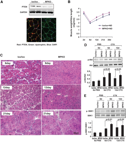
Muscle regeneration was induced by cardiotoxin injury, and we evaluated satellite cell activation and muscle maturation in HFD-fed mice. We also measured PIP(3) and the enzymes regulating its level, IRS-1-associated phosphatidylinositol 3-kinase (PI3K) and PTEN. Using primary cultures of muscle, we examined how fatty acids affect PTEN expression and how PTEN knockout influences muscle growth. Mice with muscle-specific PTEN knockout were used to examine how the HFD changes muscle regeneration.
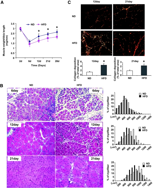
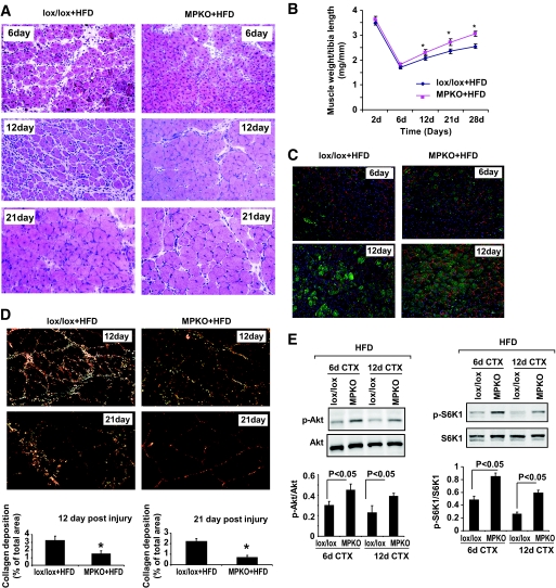
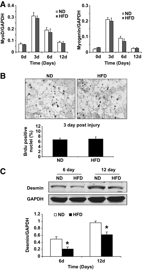
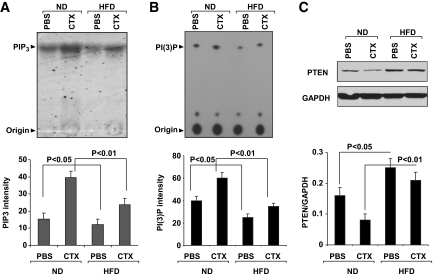
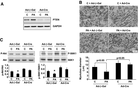
The HFD raised circulating fatty acids and impaired the growth of regenerating myofibers while delaying myofiber maturation and increasing collagen deposition. These changes were independent of impaired proliferation of muscle progenitor or satellite cells but were principally related to increased expression of PTEN, which reduced PIP(3) in muscle. In cultured muscle cells, palmitate directly stimulated PTEN expression and reduced cell growth. Knocking out PTEN restored cell growth. In mice, muscle-specific PTEN knockout improved the defects in muscle repair induced by HFD.
Insulin resistance impairs muscle regeneration by preventing myofiber maturation. The mechanism involves fatty acid-stimulated PTEN expression, which lowers muscle PIP(3). If similar pathways occur in diabetic patients, therapeutic strategies directed at improving the repair of damaged muscle could include suppression of PTEN activity.
糖尿病中损害伤口愈合的机制尚未完全阐明。为了确定这些机制,我们通过长期给予高脂肪饮食(HFD)诱导小鼠胰岛素抵抗。我们还研究了肌再生过程中磷脂酰肌醇 3,4,5-三磷酸(PIP(3))的调节,因为增强 IGF-1 信号可以改善肌肉再生。
通过心脏毒素损伤诱导肌肉再生,我们评估了 HFD 喂养小鼠中卫星细胞的激活和肌肉成熟情况。我们还测量了 PIP(3)和调节其水平的酶,胰岛素受体底物 1 相关的磷酸肌醇 3-激酶(PI3K)和 PTEN。使用肌肉原代培养物,我们研究了脂肪酸如何影响 PTEN 的表达,以及 PTEN 缺失如何影响肌肉生长。使用肌肉特异性 PTEN 缺失小鼠来研究 HFD 如何改变肌肉再生。
HFD 升高了循环脂肪酸,损害了再生肌纤维的生长,同时延迟了肌纤维成熟并增加了胶原蛋白沉积。这些变化与肌肉祖细胞或卫星细胞增殖受损无关,但主要与 PTEN 表达增加有关,这降低了肌肉中的 PIP(3)。在培养的肌肉细胞中,棕榈酸直接刺激 PTEN 表达并降低细胞生长。敲除 PTEN 恢复了细胞生长。在小鼠中,肌肉特异性 PTEN 缺失改善了 HFD 引起的肌肉修复缺陷。
胰岛素抵抗通过阻止肌纤维成熟来损害肌肉再生。该机制涉及脂肪酸刺激的 PTEN 表达,从而降低肌肉 PIP(3)。如果糖尿病患者中存在类似的途径,那么旨在改善受损肌肉修复的治疗策略可能包括抑制 PTEN 活性。