Smith Maxwell, Lucia M Scott, Werahera Priya N, La Rosa Francisco G
University of Colorado Denver, Department of Pathology, Aurora, Colorado 80045-0508, USA.
J Med Case Rep. 2010 Jan 20;4:16. doi: 10.1186/1752-1947-4-16.
Urethral carcinoid tumors are very rare tumors with only four cases described in the literature.
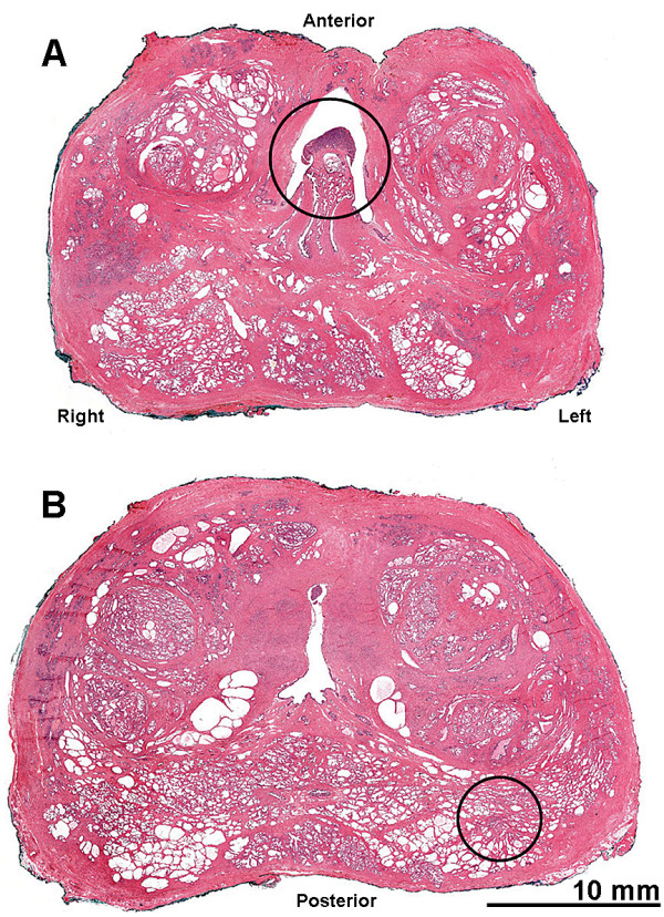
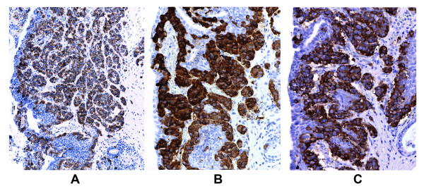
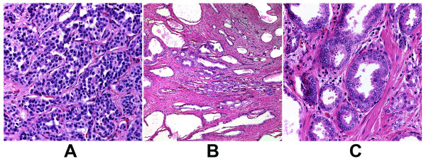
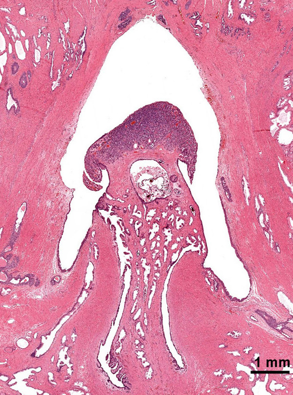
We present the case of a 61-year-old man with a primary carcinoid tumor of the verumontanum (colliculis seminalis) portion of the prostatic urethra with a coexisting prostatic adenocarcinoma. In addition to whole mount hematoxylin and eosin staining, special immunoperoxidase staining specific for chromogranin A, neuron specific enolase, synaptophysin, pan-cytokeratin and PSA, and a special combined staining for racemase (alpha-methyl CoA) antigen and p63 antigen were performed. A review of the literature is included.A single focus of invasive prostatic adenocarcinoma was identified in the periphery of the mid-left, posterior quadrant of the prostate. Approximately 17 mm from this adenocarcinoma, within the verumontanum of the prostatic urethra, there was a 3 mm maximal dimension carcinoid tumor.
Based on different histological features and antigenic profiles, we concluded that the two tumors were distinct.
尿道类癌肿瘤极为罕见,文献中仅描述了4例。
我们报告一例61岁男性,其前列腺尿道精阜(精丘)部位存在原发性类癌肿瘤,并伴有前列腺腺癌。除了进行全层苏木精和伊红染色外,还进行了针对嗜铬粒蛋白A、神经元特异性烯醇化酶、突触素、全细胞角蛋白和前列腺特异性抗原的特殊免疫过氧化物酶染色,以及针对消旋酶(α-甲基辅酶A)抗原和p63抗原的特殊联合染色。本文还包括文献综述。在前列腺左中后象限的外周发现了一个浸润性前列腺腺癌单发病灶。在距该腺癌约17毫米处,前列腺尿道的精阜内有一个最大直径为3毫米的类癌肿瘤。
基于不同的组织学特征和抗原谱,我们得出结论认为这两种肿瘤是不同的。