Rasmussen Anne S, Lauridsen Henrik, Laustsen Christoffer, Jensen Bjarke G, Pedersen Steen F, Uhrenholt Lars, Boel Lene W T, Uldbjerg Niels, Wang Tobias, Pedersen Michael
MR-research Centre, Aarhus University Hospital, Aarhus, Denmark.
BMC Physiol. 2010 Mar 12;10:3. doi: 10.1186/1472-6793-10-3.
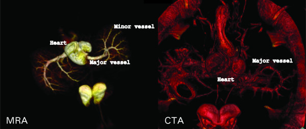
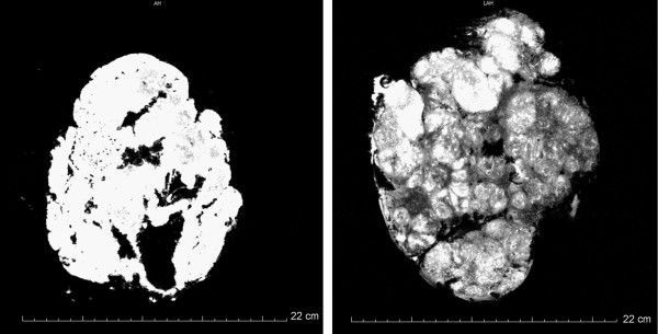
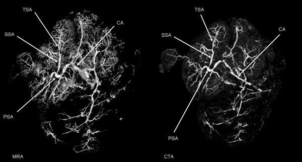
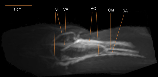
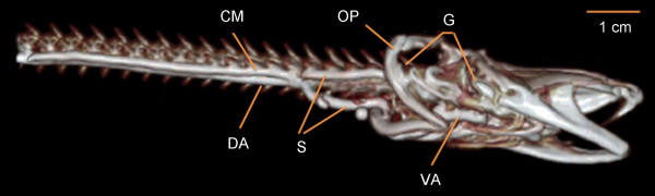
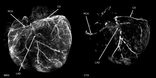
In biomedical sciences, ex vivo angiography is a practical mean to elucidate vascular structures three-dimensionally with simultaneous estimation of intravascular volume. The objectives of this study were to develop a magnetic resonance (MR) method for ex vivo angiography and to compare the findings with computed tomography (CT). To demonstrate the usefulness of this method, examples are provided from four different tissues and species: the human placenta, a rice field eel, a porcine heart and a turtle.
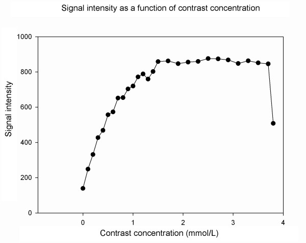
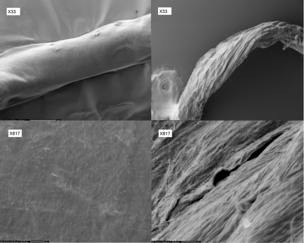
The optimal solution for ex vivo MR angiography (MRA) was a compound containing gelatine (0.05 g/mL), the CT contrast agent barium sulphate (0.43 mol/L) and the MR contrast agent gadoteric acid (2.5 mmol/L). It was possible to perform angiography on all specimens. We found that ex vivo MRA could only be performed on fresh tissue because formalin fixation makes the blood vessels permeable to the MR contrast agent.
Ex vivo MRA provides high-resolution images of fresh tissue and delineates fine structures that we were unable to visualise by CT. We found that MRA provided detailed information similar to or better than conventional CTA in its ability to visualize vessel configuration while avoiding interfering signals from adjacent bones. Interestingly, we found that vascular tissue becomes leaky when formalin-fixed, leading to increased permeability and extravascular leakage of MR contrast agent.
在生物医学科学领域,离体血管造影术是一种通过同时估计血管内体积来三维阐明血管结构的实用方法。本研究的目的是开发一种用于离体血管造影的磁共振(MR)方法,并将结果与计算机断层扫描(CT)进行比较。为了证明该方法的实用性,提供了来自四种不同组织和物种的实例:人类胎盘、稻田鳗鱼、猪心脏和乌龟。
离体磁共振血管造影(MRA)的最佳溶液是一种含有明胶(0.05 g/mL)、CT造影剂硫酸钡(0.43 mol/L)和MR造影剂钆特酸(2.5 mmol/L)的化合物。对所有标本进行血管造影都是可行的。我们发现离体MRA只能在新鲜组织上进行,因为福尔马林固定会使血管对MR造影剂具有通透性。
离体MRA可提供新鲜组织的高分辨率图像,并描绘出我们无法通过CT可视化的精细结构。我们发现,MRA在可视化血管形态方面提供了与传统CTA相似或更好的详细信息,同时避免了来自相邻骨骼的干扰信号。有趣的是,我们发现福尔马林固定后血管组织会变得渗漏,导致MR造影剂的通透性增加和血管外渗漏。