• 文献检索
  • 文档翻译
  • 深度研究
  • 学术资讯
  • Suppr Zotero 插件Zotero 插件
  • 邀请有礼
  • 套餐&价格
  • 历史记录
应用&插件
Suppr Zotero 插件Zotero 插件浏览器插件Mac 客户端Windows 客户端微信小程序
定价
高级版会员购买积分包购买API积分包
服务
文献检索文档翻译深度研究API 文档MCP 服务
关于我们
关于 Suppr公司介绍联系我们用户协议隐私条款
关注我们

Suppr 超能文献

核心技术专利:CN118964589B侵权必究
粤ICP备2023148730 号-1Suppr @ 2026

文献检索

告别复杂PubMed语法,用中文像聊天一样搜索,搜遍4000万医学文献。AI智能推荐,让科研检索更轻松。

立即免费搜索

文件翻译

保留排版,准确专业,支持PDF/Word/PPT等文件格式,支持 12+语言互译。

免费翻译文档

深度研究

AI帮你快速写综述,25分钟生成高质量综述,智能提取关键信息,辅助科研写作。

立即免费体验

64 层螺旋 CT 冠状动脉成像诊断支架内再狭窄的准确性:荟萃分析。

Diagnostic accuracy of 64-slice computed tomography coronary angiography for the detection of in-stent restenosis: a meta-analysis.

机构信息

Division of Cardiology, Careggi Hospital, V.le Morgagni, 85 Florence, Italy.

出版信息

J Nucl Cardiol. 2010 Jun;17(3):470-8. doi: 10.1007/s12350-010-9218-2. Epub 2010 Apr 9.

DOI:10.1007/s12350-010-9218-2
PMID:20379863
原文链接:https://pmc.ncbi.nlm.nih.gov/articles/PMC2866963/
Abstract

BACKGROUND

We sought to evaluate the diagnostic accuracy of 64-slice multi-detector row computed tomography (MDCT) compared with invasive coronary angiography for in-stent restenosis (ISR) detection.

METHODS

MEDLINE, Cochrane library, and BioMed Central database searches were performed until April 2009 for original articles. Inclusion criteria were (1) 64-MDCT was used as a diagnostic test for ISR, with >50% diameter stenosis selected as the cut-off criterion for significant ISR, using invasive coronary angiography and quantitative coronary angiography as the standard of reference; (2) absolute numbers of true positive, false positive, true negative, and false negative results could be derived. Standard meta-analytic methods were applied.

RESULTS

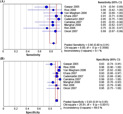
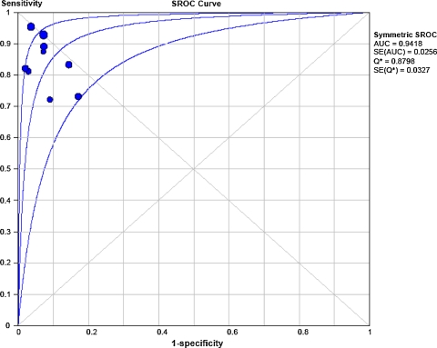
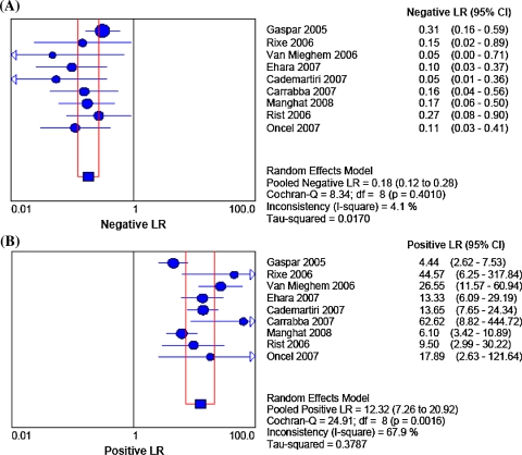
Nine studies with a total of 598 patients with 978 stents included were considered eligible. On average, 9% of stents were unassessable (range 0-42%). Accuracy tests with 95% confidence intervals (CIs) comparing 64-MDCT vs invasive coronary angiography showed that pooled sensitivity, specificity, positive and negative likelihood ratio (random effect model) values were: 86% (95% CI 80-91%), 93% (95% CI 91-95%), 12.32 (95% CI 7.26-20.92), 0.18 (95% CI 0.12-0.28) for binary ISR detection. The symmetric area under the curve value was 0.94, indicating good agreement between 64-MDCT and invasive coronary angiography.

CONCLUSIONS

64-MDCT has a good diagnostic accuracy for ISR detection with a particularly high negative predictive value. However, still a relatively large proportion of stents remains uninterpretable. Accordingly, only in selected patients, 64-MDCT may serve as a potential alternative noninvasive method to rule out ISR.

摘要

背景

我们旨在评估 64 层多排螺旋 CT(MDCT)与有创冠状动脉造影相比在检测支架内再狭窄(ISR)方面的诊断准确性。

方法

对截至 2009 年 4 月的原始文献进行了 MEDLINE、Cochrane 图书馆和 BioMed Central 数据库检索。纳入标准为:(1)64-MDCT 作为 ISR 的诊断检测方法,以 >50%的直径狭窄为显著 ISR 的截断标准,以有创冠状动脉造影和定量冠状动脉造影为参考标准;(2)可获得真阳性、假阳性、真阴性和假阴性的绝对例数。采用标准的荟萃分析方法。

结果

共纳入 9 项研究,共计 598 例患者,978 个支架。平均有 9%的支架无法评估(范围为 0-42%)。比较 64-MDCT 与有创冠状动脉造影的准确率检验显示,64-MDCT 与有创冠状动脉造影的合并敏感度、特异度、阳性和阴性似然比(随机效应模型)值分别为 86%(95% CI 80-91%)、93%(95% CI 91-95%)、12.32(95% CI 7.26-20.92)和 0.18(95% CI 0.12-0.28),用于二元 ISR 检测。曲线下面积的对称值为 0.94,表明 64-MDCT 与有创冠状动脉造影之间有良好的一致性。

结论

64-MDCT 对 ISR 的检测具有良好的诊断准确性,阴性预测值特别高。然而,仍有相当大比例的支架无法解释。因此,只有在选择的患者中,64-MDCT 可能作为一种潜在的替代非侵入性方法,用于排除 ISR。

https://cdn.ncbi.nlm.nih.gov/pmc/blobs/a32b/2866963/55a20b70784d/12350_2010_9218_Fig3_HTML.jpg
https://cdn.ncbi.nlm.nih.gov/pmc/blobs/a32b/2866963/3ed23efd2c80/12350_2010_9218_Fig1_HTML.jpg
https://cdn.ncbi.nlm.nih.gov/pmc/blobs/a32b/2866963/34ae56b1099c/12350_2010_9218_Fig2_HTML.jpg
https://cdn.ncbi.nlm.nih.gov/pmc/blobs/a32b/2866963/55a20b70784d/12350_2010_9218_Fig3_HTML.jpg
https://cdn.ncbi.nlm.nih.gov/pmc/blobs/a32b/2866963/3ed23efd2c80/12350_2010_9218_Fig1_HTML.jpg
https://cdn.ncbi.nlm.nih.gov/pmc/blobs/a32b/2866963/34ae56b1099c/12350_2010_9218_Fig2_HTML.jpg
https://cdn.ncbi.nlm.nih.gov/pmc/blobs/a32b/2866963/55a20b70784d/12350_2010_9218_Fig3_HTML.jpg

相似文献

1
Diagnostic accuracy of 64-slice computed tomography coronary angiography for the detection of in-stent restenosis: a meta-analysis.64 层螺旋 CT 冠状动脉成像诊断支架内再狭窄的准确性:荟萃分析。
J Nucl Cardiol. 2010 Jun;17(3):470-8. doi: 10.1007/s12350-010-9218-2. Epub 2010 Apr 9.
2
Diagnostic accuracy of 64-slice multidetector CT angiography for detection of in-stent restenosis of vertebral artery ostium stents: comparison with conventional angiography.64层螺旋CT血管造影术检测椎动脉开口支架内再狭窄的诊断准确性:与传统血管造影术的比较
Acta Radiol. 2014 Oct;55(8):1000-7. doi: 10.1177/0284185113507922. Epub 2013 Oct 9.
3
Multi-detector row CT angiography in the assessment of coronary in-stent restenosis: a systematic review.多排探测器CT血管造影术在评估冠状动脉支架内再狭窄中的应用:一项系统评价
Eur J Radiol. 2009 Mar;69(3):489-95. doi: 10.1016/j.ejrad.2007.11.030. Epub 2007 Dec 26.
4
Comparison of feasibility and diagnostic accuracy of 64-slice multidetector computed tomographic coronary angiography versus invasive coronary angiography versus intravascular ultrasound for evaluation of in-stent restenosis.64层螺旋计算机断层扫描冠状动脉造影术、有创冠状动脉造影术及血管内超声评估支架内再狭窄的可行性及诊断准确性比较
Am J Cardiol. 2009 May 15;103(10):1349-58. doi: 10.1016/j.amjcard.2009.01.343. Epub 2009 Mar 25.
5
Diagnostic accuracy of coronary in-stent restenosis using 64-slice computed tomography: comparison with invasive coronary angiography.64层计算机断层扫描对冠状动脉支架内再狭窄的诊断准确性:与有创冠状动脉造影的比较。
J Am Coll Cardiol. 2007 Mar 6;49(9):951-9. doi: 10.1016/j.jacc.2006.10.065. Epub 2007 Feb 20.
6
In-stent area stenosis on 64-slice multi-detector computed tomography coronary angiography: optimal cutoff value for minimum lumen cross-sectional area of coronary stents compared with intravascular ultrasound.64 层多排螺旋 CT 冠状动脉造影显示支架内面积狭窄:支架最小管腔横截面积的最佳截断值与血管内超声比较。
Int J Cardiovasc Imaging. 2012 Jun;28 Suppl 1:21-31. doi: 10.1007/s10554-012-0057-x. Epub 2012 May 6.
7
Coronary in-stent restenosis: predisposing clinical and stent-related factors, diagnostic performance and analyses of inaccuracies in 320-row computed tomography angiography.冠状动脉支架内再狭窄:相关临床及支架因素、诊断性能以及320排计算机断层扫描血管造影术的误差分析
Int J Cardiovasc Imaging. 2016 Jun;32 Suppl 1:105-15. doi: 10.1007/s10554-016-0872-6. Epub 2016 Apr 22.
8
[Diagnostic value of 320-slice computed tomography coronary angiography to assess in-stent restenosis].320层螺旋CT冠状动脉血管造影对评估支架内再狭窄的诊断价值
Zhonghua Xin Xue Guan Bing Za Zhi. 2012 Jun;40(6):487-91.
9
Usefulness of 64-slice multidetector computed tomography for detecting drug eluting in-stent restenosis.64层螺旋CT在检测药物洗脱支架内再狭窄中的应用价值
Am J Cardiol. 2007 Dec 15;100(12):1754-8. doi: 10.1016/j.amjcard.2007.07.038. Epub 2007 Oct 29.
10
Diagnostic accuracy of 320-row multidetector computed tomography coronary angiography to noninvasively assess in-stent restenosis.320 排多层螺旋 CT 冠状动脉成像无创评估支架内再狭窄的诊断准确性。
Invest Radiol. 2010 Jun;45(6):331-40. doi: 10.1097/RLI.0b013e3181dfa312.

引用本文的文献

1
Cardiac Repair and Regeneration via Advanced Technology: Narrative Literature Review.通过先进技术实现心脏修复与再生:叙述性文献综述
JMIR Biomed Eng. 2025 Mar 8;10:e65366. doi: 10.2196/65366.
2
Cardiovascular Computed Tomography and Magnetic Resonance Imaging Guideline of the Brazilian Society of Cardiology and the Brazilian College of Radiology - 2024.巴西心脏病学会和巴西放射学会心血管计算机断层扫描与磁共振成像指南 - 2024年
Arq Bras Cardiol. 2024 Oct 28;121(9):e20240608. doi: 10.36660/abc.20240608.
3
Effect of deep learning image reconstruction with high-definition standard scan mode on image quality of coronary stents and arteries.

本文引用的文献

1
Diagnostic accuracy of 320-row multidetector computed tomography coronary angiography to noninvasively assess in-stent restenosis.320 排多层螺旋 CT 冠状动脉成像无创评估支架内再狭窄的诊断准确性。
Invest Radiol. 2010 Jun;45(6):331-40. doi: 10.1097/RLI.0b013e3181dfa312.
2
High-definition multidetector computed tomography for evaluation of coronary artery stents: comparison to standard-definition 64-detector row computed tomography.用于评估冠状动脉支架的高清多排螺旋计算机断层扫描:与标准清晰度64排螺旋计算机断层扫描的比较
J Cardiovasc Comput Tomogr. 2009 Jul-Aug;3(4):246-51. doi: 10.1016/j.jcct.2009.06.006. Epub 2009 Jun 12.
3
深度学习图像重建结合高清标准扫描模式对冠状动脉支架及血管图像质量的影响
Quant Imaging Med Surg. 2024 Feb 1;14(2):1616-1635. doi: 10.21037/qims-23-1064. Epub 2024 Jan 17.
4
Evolving Coronary Stent Technologies - A Glimpse Into the Future.不断发展的冠状动脉支架技术——展望未来
Cureus. 2023 Mar 1;15(3):e35651. doi: 10.7759/cureus.35651. eCollection 2023 Mar.
5
Clinical experience regarding the diagnostic value of segment-by-segment coronary computed tomography angiography in comparison with that of invasive coronary angiography.与有创冠状动脉造影相比,冠状动脉计算机断层扫描血管造影逐段分析的诊断价值的临床经验。
Radiol Bras. 2022 May-Jun;55(3):156-160. doi: 10.1590/0100-3984.2021.0092.
6
Added Value of Computed Tomography Virtual Intravascular Endoscopy in the Evaluation of Coronary Arteries with Stents or Plaques.计算机断层扫描虚拟血管内内窥镜检查在评估有支架或斑块的冠状动脉中的附加价值。
Diagnostics (Basel). 2022 Feb 3;12(2):390. doi: 10.3390/diagnostics12020390.
7
Coronary Computer Tomography Angiography in 2021-Acquisition Protocols, Tips and Tricks and Heading beyond the Possible.2021年冠状动脉计算机断层扫描血管造影——采集方案、技巧及超越可能的进展
Diagnostics (Basel). 2021 Jun 10;11(6):1072. doi: 10.3390/diagnostics11061072.
8
Stent evaluation by coronary computed tomography angiography: a comparison between Iopamidol-370 and Ioversol-320 hypo-osmolar iodine concentration contrasts.冠状动脉计算机断层血管造影术评估支架:碘帕醇 370 与碘佛醇 320 低渗碘浓度对比剂的比较。
Br J Radiol. 2020 Nov 1;93(1115):20200078. doi: 10.1259/bjr.20200078. Epub 2020 Aug 20.
9
Health Care Monitoring and Treatment for Coronary Artery Diseases: Challenges and Issues.冠状动脉疾病的医疗监测与治疗:挑战与问题。
Sensors (Basel). 2020 Aug 1;20(15):4303. doi: 10.3390/s20154303.
10
Functional and Anatomical Imaging in Patients with Ischemic Symptoms and Known Coronary Artery Disease.有缺血症状和已知冠心病患者的功能和解剖成像。
Curr Cardiol Rep. 2019 Jul 1;21(8):79. doi: 10.1007/s11886-019-1155-3.
Diagnostic accuracy of 64 multislice CT angiography in the assessment of coronary in-stent restenosis: a meta-analysis.
64 层多层螺旋 CT 血管造影术评估冠状动脉支架内再狭窄的诊断准确性:荟萃分析。
Eur J Radiol. 2010 Feb;73(2):266-73. doi: 10.1016/j.ejrad.2008.10.025. Epub 2008 Dec 3.
4
Dual-source CT in step-and-shoot mode: noninvasive coronary angiography with low radiation dose.步进式双源CT:低辐射剂量的无创冠状动脉造影
Radiology. 2008 Oct;249(1):71-80. doi: 10.1148/radiol.2483072032.
5
Usefulness of 64-detector row computed tomography for evaluation of intracoronary stents in symptomatic patients with suspected in-stent restenosis.64排螺旋CT对有症状的疑似支架内再狭窄患者冠状动脉支架评估的应用价值
Am J Cardiol. 2008 Jun 1;101(11):1567-73. doi: 10.1016/j.amjcard.2008.01.042. Epub 2008 Apr 2.
6
Usefulness of 64-slice multidetector computed tomography for detecting drug eluting in-stent restenosis.64层螺旋CT在检测药物洗脱支架内再狭窄中的应用价值
Am J Cardiol. 2007 Dec 15;100(12):1754-8. doi: 10.1016/j.amjcard.2007.07.038. Epub 2007 Oct 29.
7
Dual source coronary computed tomography angiography for detecting in-stent restenosis.双源冠状动脉计算机断层扫描血管造影术用于检测支架内再狭窄
Heart. 2008 Jul;94(7):848-54. doi: 10.1136/hrt.2007.126474. Epub 2007 Sep 19.
8
Diagnostic accuracy of in-stent coronary restenosis detection with multislice spiral computed tomography: a meta-analysis.多层螺旋计算机断层扫描检测冠状动脉支架内再狭窄的诊断准确性:一项荟萃分析。
Eur Radiol. 2008 Feb;18(2):217-25. doi: 10.1007/s00330-007-0743-6. Epub 2007 Sep 1.
9
Usefulness of 64-slice multislice computed tomography coronary angiography to assess in-stent restenosis.64层多层螺旋CT冠状动脉造影评估支架内再狭窄的效用。
J Am Coll Cardiol. 2007 Jun 5;49(22):2204-10. doi: 10.1016/j.jacc.2007.02.045. Epub 2007 May 23.
10
Diagnostic accuracy of coronary in-stent restenosis using 64-slice computed tomography: comparison with invasive coronary angiography.64层计算机断层扫描对冠状动脉支架内再狭窄的诊断准确性:与有创冠状动脉造影的比较。
J Am Coll Cardiol. 2007 Mar 6;49(9):951-9. doi: 10.1016/j.jacc.2006.10.065. Epub 2007 Feb 20.