Department of Neurology and the Stroke and Cerebrovascular Center, Samsung Medical Center, Sungkyunkwan University School of Medicine, Seoul, Korea.
J Clin Neurol. 2010 Mar;6(1):41-5. doi: 10.3988/jcn.2010.6.1.41. Epub 2010 Mar 26.
It has recently been suggested that diffusion and perfusion MRI can identify subgroups likely to benefit or potentially be harmed by reperfusion therapies.
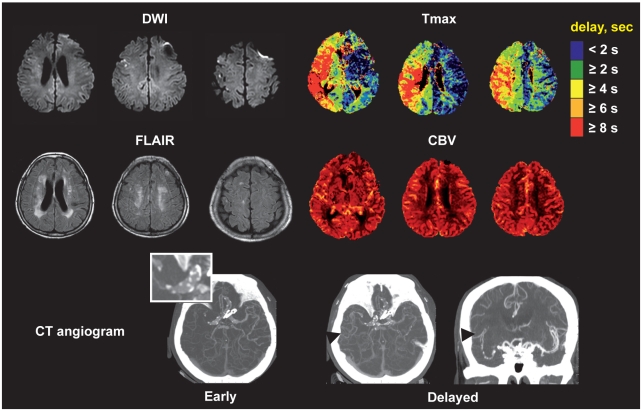
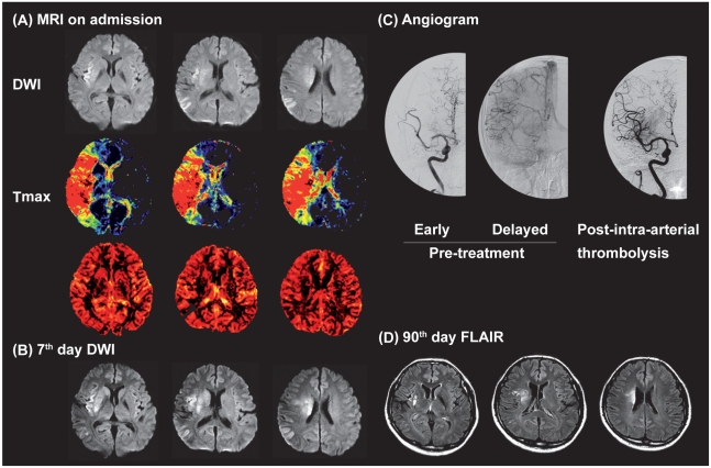
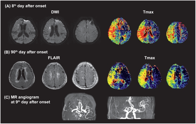
We investigated serial MRI data of two patients with occlusion of the proximal middle cerebral artery (MCA). In both cases, acute multiple cortical infarcts evident on diffusion-weighted imaging (DWI) and perfusion-weighted imaging (PWI) showed extensive areas of severe perfusion delays, indicating a malignant MRI profile. However, despite the malignant MRI profiles in these cases, no new ischemic lesions or hemorrhage evolved even in the presence of persistent arterial occlusion, and the patients recovered without sequelae.
These two cases suggest that time-domain PWI findings should be interpreted with caution in certain scenarios of acute ischemic stroke.
最近有人提出,弥散和灌注 MRI 可以识别可能受益或可能受到再灌注治疗损害的亚组。
我们研究了两名大脑中动脉近端闭塞患者的连续 MRI 数据。在这两种情况下,弥散加权成像 (DWI) 和灌注加权成像 (PWI) 显示的急性多发性皮质梗死均显示出广泛的严重灌注延迟区域,表明 MRI 表现为恶性。然而,尽管这两种情况的 MRI 表现均为恶性,但即使在动脉持续闭塞的情况下,也没有新的缺血性病灶或出血演变,且患者恢复后无后遗症。
这两个病例表明,在急性缺血性脑卒中的某些情况下,应谨慎解读时间域 PWI 结果。