• 文献检索
  • 文档翻译
  • 深度研究
  • 学术资讯
  • Suppr Zotero 插件Zotero 插件
  • 邀请有礼
  • 套餐&价格
  • 历史记录
应用&插件
Suppr Zotero 插件Zotero 插件浏览器插件Mac 客户端Windows 客户端微信小程序
定价
高级版会员购买积分包购买API积分包
服务
文献检索文档翻译深度研究API 文档MCP 服务
关于我们
关于 Suppr公司介绍联系我们用户协议隐私条款
关注我们

Suppr 超能文献

核心技术专利:CN118964589B侵权必究
粤ICP备2023148730 号-1Suppr @ 2026

文献检索

告别复杂PubMed语法,用中文像聊天一样搜索,搜遍4000万医学文献。AI智能推荐,让科研检索更轻松。

立即免费搜索

文件翻译

保留排版,准确专业,支持PDF/Word/PPT等文件格式,支持 12+语言互译。

免费翻译文档

深度研究

AI帮你快速写综述,25分钟生成高质量综述,智能提取关键信息,辅助科研写作。

立即免费体验

肾细胞癌伴恶性腹水的细胞形态学分析:2例报告

Cytomorphologic consideration in malignant ascites with renal cell carcinoma: A report of two cases.

作者信息

Gupta Ruchika, Mathur Sandeep R, Iyer Venkateswaran K, Kumar A Sudheer, Seth Amlesh

机构信息

Departments of Pathology & Urology, All India Institute of Medical Sciences, New Delhi, India.

出版信息

Cytojournal. 2010 Apr 6;7:4. doi: 10.4103/1742-6413.62256.

DOI:10.4103/1742-6413.62256
PMID:20436788
原文链接:https://pmc.ncbi.nlm.nih.gov/articles/PMC2861821/
Abstract

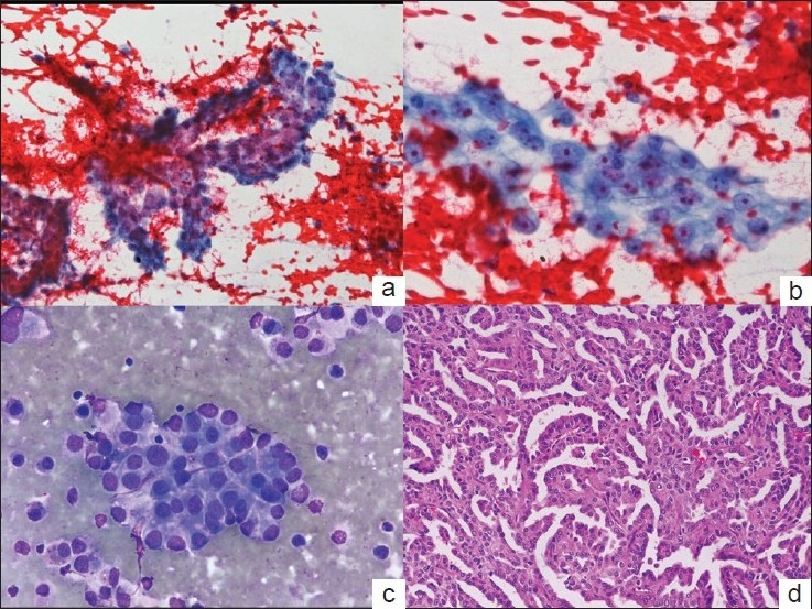
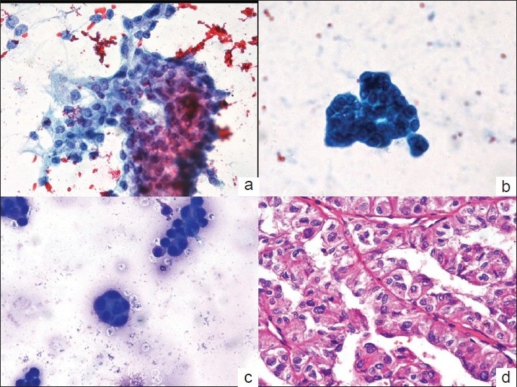
Effusions, especially peritoneal, are seen in less than 2% of patients with renal cell carcinoma (RCC). Since the tumor cells in RCC are bland and nondescript, the involvement of serous effusions is difficult to diagnose. An accurate recognition of malignant effusion and differentiation from reactive mesothelial cells is imperative. A 55-year-old male presented with gradually progressive ascites. Cytospin preparations from ascitic fluid showed reactive mesothelial cells admixed with few smooth-contoured clusters of cells with moderate cytoplasm, vesicular nuclei with prominent nucleolus. He had undergone nephrectomy for papillary RCC two years earlier. Another 36-year-old man underwent left nephrectomy for suspected RCC. Intra-operative ascitic fluid was sent for cytologic examination and showed numerous reactive mesothelial cells along with few clusters of cells with scant to moderate amount of cytoplasm, vesicular nucleus and a small nucleolus. Considering the histomorphology of the primary renal tumor in both cases, a cytologic diagnosis of malignant peritoneal effusion, morphologically compatible with RCC was rendered. RCC, due to its bland cytologic features, is easily overlooked in effusions. In a known patient, the cytopathologist must be extra vigilant to pick up the few cell clusters present in the fluid preparations and differentiate them from reactive mesothelial cells. A close inspection of the cytologic features and comparison with the histopathology of the primary tumor helps in making an accurate diagnosis.

摘要

积液,尤其是腹腔积液,在不到2%的肾细胞癌(RCC)患者中可见。由于RCC中的肿瘤细胞形态温和且无特征性,浆液性积液的累及难以诊断。准确识别恶性积液并与反应性间皮细胞进行鉴别至关重要。一名55岁男性出现逐渐加重的腹水。腹水的细胞离心涂片显示反应性间皮细胞与少数轮廓光滑的细胞簇混合,这些细胞簇具有中等量的细胞质、泡状核和明显的核仁。他两年前因乳头状RCC接受了肾切除术。另一名36岁男性因疑似RCC接受了左肾切除术。术中腹水送检进行细胞学检查,显示大量反应性间皮细胞以及少数细胞簇,这些细胞簇的细胞质含量少至中等、核呈泡状且有小核仁。考虑到这两个病例中原发性肾肿瘤的组织形态学,做出了恶性腹腔积液的细胞学诊断,形态学上与RCC相符。由于RCC的细胞特征温和,在积液中很容易被忽视。对于已知患者,细胞病理学家必须格外警惕,以识别液体制片中存在的少数细胞簇,并将它们与反应性间皮细胞区分开来。仔细检查细胞学特征并与原发性肿瘤的组织病理学进行比较有助于做出准确诊断。

https://cdn.ncbi.nlm.nih.gov/pmc/blobs/994b/2861821/81b137ca80dc/CJ-07-4-g002.jpg
https://cdn.ncbi.nlm.nih.gov/pmc/blobs/994b/2861821/6d032a9ea5b0/CJ-07-4-g001.jpg
https://cdn.ncbi.nlm.nih.gov/pmc/blobs/994b/2861821/81b137ca80dc/CJ-07-4-g002.jpg
https://cdn.ncbi.nlm.nih.gov/pmc/blobs/994b/2861821/6d032a9ea5b0/CJ-07-4-g001.jpg
https://cdn.ncbi.nlm.nih.gov/pmc/blobs/994b/2861821/81b137ca80dc/CJ-07-4-g002.jpg

相似文献

1
Cytomorphologic consideration in malignant ascites with renal cell carcinoma: A report of two cases.肾细胞癌伴恶性腹水的细胞形态学分析:2例报告
Cytojournal. 2010 Apr 6;7:4. doi: 10.4103/1742-6413.62256.
2
Small orangiophilic squamous-like cells: an underrecognized and useful morphological feature for the diagnosis of malignant mesothelioma in pleural effusion cytology.小型嗜橘红色类鳞状细胞:胸腔积液细胞学中诊断恶性间皮瘤的一种被低估但有用的形态学特征。
Cancer Cytopathol. 2014 Jan;122(1):70-5. doi: 10.1002/cncy.21345. Epub 2013 Aug 26.
3
Serous surface carcinoma of the peritoneum: useful role of cytology in differential diagnosis and follow-up.
Acta Cytol. 1996 May-Jun;40(3):429-36. doi: 10.1159/000333894.
4
Immunohistochemistry for the detection of renal cell carcinoma in effusion cytology.免疫组织化学在积液细胞学中用于检测肾细胞癌。
Diagn Cytopathol. 2011 Feb;39(2):118-23. doi: 10.1002/dc.21375.
5
Diagnostic usefulness of EMA, IMP3, and GLUT-1 for the immunocytochemical distinction of malignant cells from reactive mesothelial cells in effusion cytology using cytospin preparations.在利用细胞离心涂片制备的积液细胞学检查中,EMA、IMP3和GLUT-1在免疫细胞化学鉴别恶性细胞与反应性间皮细胞方面的诊断价值。
Diagn Cytopathol. 2011 Jun;39(6):395-401. doi: 10.1002/dc.21398. Epub 2010 May 27.
6
Approach to Diagnostic Cytopathology of Serous Effusions.浆液性积液的诊断细胞病理学方法
Cytojournal. 2021 Dec 6;18:32. doi: 10.25259/CMAS_02_03_2021. eCollection 2021.
7
Cytologic re-evaluation of negative effusions from patients with malignant mesothelioma.恶性间皮瘤患者阴性积液的细胞学重新评估。
Pathologica. 2011 Dec;103(6):318-24.
8
IMP3/L523S, a novel immunocytochemical marker that distinguishes benign and malignant cells: the expression profiles of IMP3/L523S in effusion cytology.IMP3/L523S,一种新型免疫细胞化学标志物,可区分良恶性细胞:IMP3/L523S 在积液细胞学中的表达谱。
Hum Pathol. 2010 May;41(5):745-50. doi: 10.1016/j.humpath.2009.04.030. Epub 2010 Jan 13.
9
Identification of adenocarcinoma in cytospin preparations of effusions using monoclonal antibody B72.3.使用单克隆抗体B72.3在积液细胞离心涂片制备物中鉴定腺癌。
Am J Clin Pathol. 1986 Jul;86(1):10-8. doi: 10.1093/ajcp/86.1.10.
10
Utility of anti-L523S antibody in the diagnosis of benign and malignant serous effusions.抗L523S抗体在良恶性浆液性积液诊断中的应用
Cancer. 2008 Feb 25;114(1):49-56. doi: 10.1002/cncr.23254.

引用本文的文献

1
Treatment of metastatic TFE3 microphthalmia transcription factor translocation renal cell carcinoma: a case report.转移性TFE3(小眼畸形转录因子)易位性肾细胞癌的治疗:一例报告
Transl Pediatr. 2024 Mar 27;13(3):499-507. doi: 10.21037/tp-24-35. Epub 2024 Mar 22.
2
Authors attain comparable or slightly higher rates of citation publishing in an open access journal (CytoJournal) compared to traditional cytopathology journals - A five year (2007-2011) experience.与传统细胞病理学期刊相比,作者在开放获取期刊(《细胞杂志》)上发表文章获得的引用率相当或略高——一项为期五年(2007 - 2011年)的经验。
Cytojournal. 2014 Apr 29;11:10. doi: 10.4103/1742-6413.131739. eCollection 2014.
3

本文引用的文献

1
Expression of renal cell carcinoma-associated markers erythropoietin, CD10, and renal cell carcinoma marker in diffuse malignant mesothelioma and metastatic renal cell carcinoma.肾细胞癌相关标志物促红细胞生成素、CD10及肾细胞癌标志物在弥漫性恶性间皮瘤和转移性肾细胞癌中的表达
Arch Pathol Lab Med. 2006 Jun;130(6):823-7. doi: 10.5858/2006-130-823-EORCCM.
2
The diagnostic utility of MOC31, BerEP4, RCC marker and CD10 in the classification of renal cell carcinoma and renal oncocytoma: an immunohistochemical analysis of 328 cases.MOC31、BerEP4、肾细胞癌标志物及CD10在肾细胞癌和肾嗜酸细胞瘤分类中的诊断效用:328例免疫组织化学分析
Histopathology. 2004 Nov;45(5):452-9. doi: 10.1111/j.1365-2559.2004.01962.x.
3
Announcement of first time Cytojournal impact factor for 2012 coincides with Cytojournal decade celebration (2004-2013).
2012年《细胞杂志》首次影响因子的公布恰逢《细胞杂志》十周年庆典(2004 - 2013年)。
Cytojournal. 2013 Aug 30;10:18. doi: 10.4103/1742-6413.117359. eCollection 2013.
4
A review of uncommon cytopathologic diagnoses of pleural effusions from a chest diseases center in Turkey.土耳其一家胸部疾病中心对胸腔积液罕见细胞病理学诊断的综述。
Cytojournal. 2011;8:13. doi: 10.4103/1742-6413.83026. Epub 2011 Jul 16.
Effusion cytology of renal cell carcinoma.
Cancer. 1998 Jun 25;84(3):148-52. doi: 10.1002/(sici)1097-0142(19980625)84:3<148::aid-cncr5>3.0.co;2-k.
4
Cytologic findings of collecting duct carcinoma of the kidney.
Diagn Cytopathol. 1995 Nov;13(4):304-9. doi: 10.1002/dc.2840130406.
5
Pleural effusion in renal cell carcinoma--a rare presenting feature.肾细胞癌中的胸腔积液——一种罕见的临床表现。
Indian J Chest Dis Allied Sci. 1987 Jan-Mar;29(1):29-35.
6
The cytologic diagnosis of malignant neoplasms in pleural and peritoneal effusions.胸膜和腹腔积液中恶性肿瘤的细胞学诊断
Acta Cytol. 1987 Mar-Apr;31(2):85-97.
7
Identification of types and primary sites of malignant tumors by examination of exfoliated tumor cells in serous fluids. Comparison with the diagnostic accuracy on small histologic biopsies.通过检查浆液性液体中的脱落肿瘤细胞来鉴定恶性肿瘤的类型和原发部位。与小组织活检的诊断准确性进行比较。
Acta Cytol. 1985 Sep-Oct;29(5):753-67.
8
Pleural effusion and renal cell carcinoma: an angiographic-pathologic correlation.胸腔积液与肾细胞癌:血管造影-病理相关性研究
Chest. 1979 May;75(5):647-8. doi: 10.1378/chest.75.5.647-b.