Department of Radiology, West China Hospital of Sichuan University, 37# Guo Xue Xiang, Chengdu, Sichuan 610041, China.
Eur J Med Res. 2010 Feb 26;15(2):84-7. doi: 10.1186/2047-783x-15-2-84.
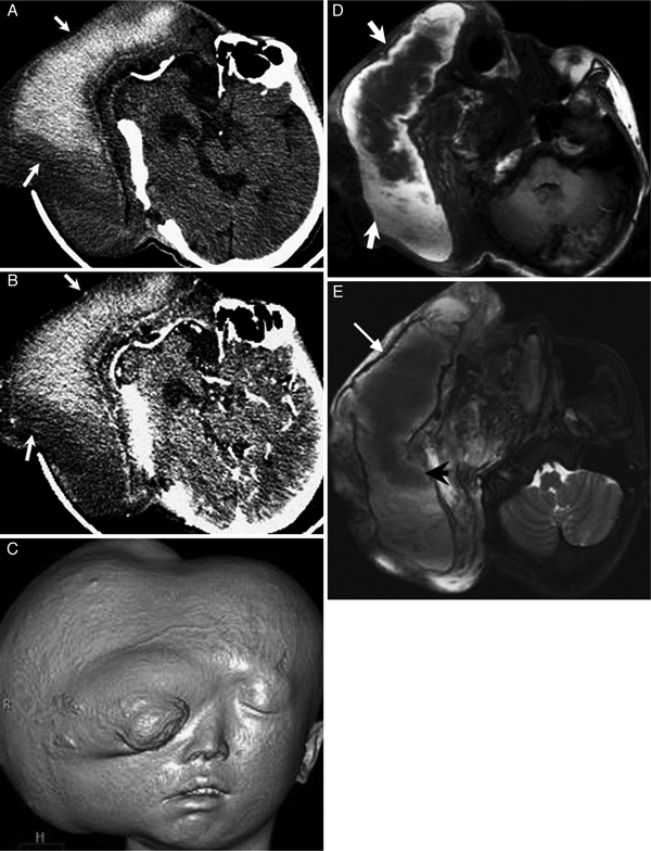
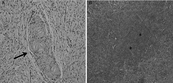
Plexiform neurofibroma (PN) is a rare benign tumor and a special subtype of neurofibromatosis type 1 (NF1). Though the incidence is low, giant PN of the craniomaxillofacial region could result in severe hemifacial hypertrophy which is known as a typical manifestation of NF1 in young children. Here, we retrospectively reported a giant plexiform neurofibroma with hemorrhage in the cranio-maxillofacial region detected by CT and MRI. In addition, a brief review of the relevant literature is presented.
丛状神经纤维瘤(PN)是一种罕见的良性肿瘤,也是神经纤维瘤病 1 型(NF1)的特殊亚型。尽管发病率较低,但发生于颅颌面区域的巨大丛状神经纤维瘤可导致严重的半侧面部肥大,这是 NF1 在幼儿中的一个典型表现。在此,我们回顾性报告了一例 CT 和 MRI 检查发现的颅颌面区域巨大丛状神经纤维瘤伴出血病例,并对相关文献进行了简要复习。