Institute of Medical Microbiology and Hygiene, University Medical Center, Johannes Gutenberg-University Mainz, Mainz, Germany.
Med Microbiol Immunol. 2010 Nov;199(4):299-309. doi: 10.1007/s00430-010-0163-0. Epub 2010 May 8.
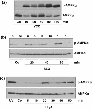
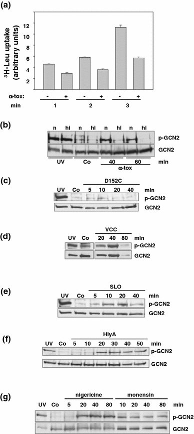
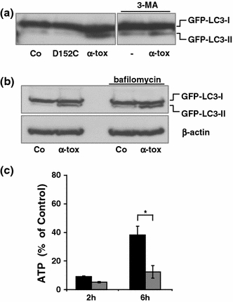
Pore-forming toxins (PFT) comprise a large, structurally heterogeneous group of bacterial protein toxins. Nucleated target cells mount complex responses which allow them to survive moderate membrane damage by PFT. Autophagy has recently been implicated in responses to various PFT, but how this process is triggered is not known, and the significance of the phenomenon is not understood. Here, we show that S. aureus α-toxin, Vibrio cholerae cytolysin, streptolysin O and E. coli haemolysin activate two pathways leading to autophagy. The first pathway is triggered via AMP-activated protein kinase (AMPK). AMPK is a major energy sensor which induces autophagy by inhibiting the target of rapamycin complex 1 (TORC1) in response to a drop of the cellular ATP/AMP-ratio, as is also observed in response to membrane perforation. The second pathway is activated by the conserved eIF2α-kinase GCN2, which causes global translational arrest and promotes autophagy in response to starvation. The latter could be accounted for by impaired amino acid transport into target cells. Notably, PKR, an eIF2α-kinase which has been implicated in autophagy induction during viral infection, was also activated upon membrane perforation, and evidence was obtained that phosphorylation of eIF2α is required for the accumulation of autophagosomes in α-toxin-treated cells. Treatment with 3-methyl-adenine inhibited autophagy and disrupted the ability of cells to recover from sublethal attack by S. aureus α-toxin. We propose that PFT induce pro-autophagic signals through membrane perforation-dependent nutrient and energy depletion, and that an important function of autophagy in this context is to maintain metabolic homoeostasis.
孔形成毒素 (PFT) 是一大组结构多样的细菌蛋白毒素。有核靶细胞会产生复杂的反应,使它们能够在中等程度的膜损伤下存活下来。自噬最近被牵连到各种 PFT 的反应中,但这个过程是如何触发的还不清楚,而且这个现象的意义也不理解。在这里,我们表明金黄色葡萄球菌α-毒素、霍乱弧菌细胞溶解素、链球菌溶素 O 和大肠杆菌溶血素激活了两条导致自噬的途径。第一条途径是通过 AMP 激活的蛋白激酶 (AMPK) 触发的。AMPK 是一种主要的能量传感器,当细胞内 ATP/AMP 比值下降时,它通过抑制雷帕霉素复合物 1 (TORC1) 来诱导自噬,这与膜穿孔时的反应也是一样的。第二条途径是由保守的 eIF2α-激酶 GCN2 激活的,它导致全球翻译抑制,并在饥饿时促进自噬。后者可能是由于氨基酸向靶细胞的转运受损所致。值得注意的是,在病毒感染过程中被牵连到自噬诱导的 eIF2α-激酶 PKR,在膜穿孔时也被激活,并且有证据表明 eIF2α 的磷酸化是 α-毒素处理细胞中自噬体积累所必需的。用 3-甲基腺嘌呤处理可抑制自噬,并破坏细胞从金黄色葡萄球菌α-毒素的亚致死攻击中恢复的能力。我们提出,PFT 通过膜穿孔依赖性的营养和能量耗竭诱导促自噬信号,而自噬在这种情况下的一个重要功能是维持代谢平衡。