Olin Neuropsychiatry Research Center, Institute of Living, Hartford Hospital, Hartford, Connecticut 06106, USA.
Biol Psychiatry. 2010 Jul 1;68(1):61-9. doi: 10.1016/j.biopsych.2010.03.035. Epub 2010 May 23.
Schizophrenia is hypothesized to involve disordered connectivity between brain regions. Currently, there are no direct measures of brain connectivity; functional and structural connectivity used separately provide only limited insight. Simultaneous measure of anatomical and functional connectivity and its interactions allow for better understanding of schizophrenia-related alternations in brain connectivity.
Twenty-seven schizophrenia patients and 27 healthy control subjects underwent magnetic resonance imaging with resting state functional magnetic resonance imaging and diffusion tensor imaging. Separate functional and anatomical connectivity maps were calculated and combined for each subject. Global, regional, and voxel measures and K-means network analysis were employed to identify group differences and correlation with clinical symptoms.
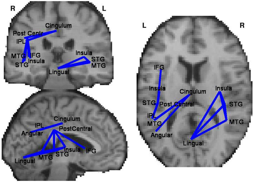
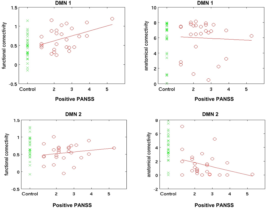
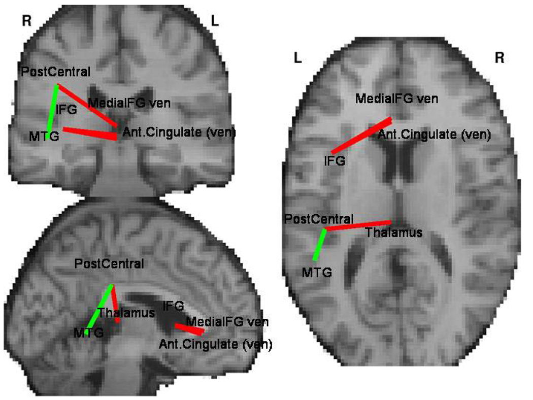
A global connectivity analysis indicated that patients had lower anatomical connectivity and lower coherence between the two imaging modalities. In schizophrenia these group differences correlated with clinical symptom severity. Although anatomical connectivity nearly uniformly decreased, functional connectivity in schizophrenia was lower for some connections (e.g., middle temporal gyrus) and higher for others (e.g., cingulate and thalamus). Within the default mode network (DMN) two separate subsystems can be identified. Schizophrenia patients showed decoupling between structural and functional connectivity that can be localized to networks originating in posterior cingulate cortex as well as in the task-positive network and one of the DMN components.
Combining two measures of brain connectivity provides more comprehensive descriptions of altered brain connectivity underlying schizophrenia. Patients show deficits in white matter anatomy, but functional connectivity alterations are more complex. Fusion of both methods allows identification of subsystems showing both increased and decreased functional connectivity.
精神分裂症被假设涉及大脑区域之间的连接紊乱。目前,没有大脑连接的直接测量方法;单独使用功能和结构连接只能提供有限的见解。同时测量解剖和功能连接及其相互作用,可以更好地理解与精神分裂症相关的大脑连接变化。
27 名精神分裂症患者和 27 名健康对照者接受了磁共振成像、静息态功能磁共振成像和弥散张量成像。为每个受试者分别计算并组合了功能和解剖连接图。采用全局、区域和体素测量以及 K-均值网络分析来识别组间差异,并与临床症状相关。
全局连接分析表明,患者的解剖连接较低,两种成像方式之间的相干性也较低。在精神分裂症中,这些组间差异与临床症状严重程度相关。尽管解剖连接几乎普遍下降,但精神分裂症患者的功能连接在某些连接(如颞中回)较低,而在其他连接(如扣带回和丘脑)较高。在默认模式网络(DMN)中,可以识别出两个独立的子系统。精神分裂症患者表现出结构和功能连接之间的解耦,这种解耦可以定位于起源于后扣带皮层的网络,以及任务正网络和 DMN 组件之一。
结合两种大脑连接的测量方法,可以更全面地描述精神分裂症相关的大脑连接变化。患者的白质解剖存在缺陷,但功能连接的改变更为复杂。两种方法的融合可以识别出显示功能连接增加和减少的子系统。