Department of Orthopaedic Surgery, Yeungnam University College of Medicine, Daegu, Korea.
Clin Orthop Surg. 2010 Jun;2(2):76-84. doi: 10.4055/cios.2010.2.2.76. Epub 2010 May 4.
This study evaluated the clinical results of arthroscopically assisted single and double bundle tibial inlay reconstructions of an isolated posterior cruciate ligament (PCL) injury.
This study reviewed the data for 14 patients who underwent a single bundle tibial inlay PCL reconstruction (Group A) and 16 patients who underwent a double bundle tibial inlay PCL reconstruction (Group B) between August 1999 and August 2002. The mean follow-up period in groups A and B was 90.5 months and 64 months, respectively.
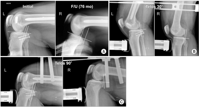
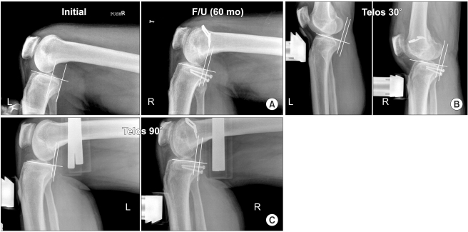
The Lysholm knee scores in groups A and B increased from an average of 43.3 +/- 7.04 and 44.7 +/- 5.02 preoperatively to 88.1 +/- 7.32 and 88.7 +/- 9.11 points at the final follow-up, respectively. In group A, stress radiography using a Telos device showed that the preoperative mean side-to-side differences (SSDs) of 9.5 +/- 1.60 mm at 30 degrees of flexion and 9.8 +/- 1.70 mm at 90 degrees of flexion were improved to 2.8 +/- 1.19 mm and 3.0 +/- 1.1 mm, respectively. In group B, the preoperative SSDs of 10.4 +/- 1.50 mm at 30 degrees of flexion and 10.7 +/- 1.60 mm at 90 degrees of flexion improved to 2.7 +/- 1.15 mm and 2.6 +/- 0.49 mm, respectively. There was no significant difference in the clinical scores and radiologic findings between the two groups.
Single bundle and double bundle PCL reconstructions using the tibial inlay technique give satisfactory clinical results in patients with an isolated PCL injury, and there are no significant differences in the clinical and radiological results between the two techniques. These results suggest that it is unnecessary to perform the more technically challenging double bundle reconstruction using the tibial inlay technique in an isolated PCL injury.
本研究评估了关节镜辅助下单独后交叉韧带(PCL)损伤的单束和双束胫骨镶嵌重建的临床结果。
本研究回顾了 1999 年 8 月至 2002 年 8 月期间接受单束胫骨镶嵌 PCL 重建(A 组)和 16 例接受双束胫骨镶嵌 PCL 重建(B 组)的 14 例患者的数据。A 组和 B 组的平均随访时间分别为 90.5 个月和 64 个月。
A 组和 B 组的 Lysholm 膝关节评分分别从术前平均 43.3 +/- 7.04 和 44.7 +/- 5.02 分增加到最终随访时的 88.1 +/- 7.32 和 88.7 +/- 9.11 分。在 A 组中,使用 Telos 设备进行的应力放射摄影显示,术前屈曲 30 度时平均侧到侧差异(SSD)为 9.5 +/- 1.60 毫米,屈曲 90 度时为 9.8 +/- 1.70 毫米,分别改善至 2.8 +/- 1.19 毫米和 3.0 +/- 1.1 毫米。在 B 组中,术前屈曲 30 度时 SSD 为 10.4 +/- 1.50 毫米,屈曲 90 度时为 10.7 +/- 1.60 毫米,分别改善至 2.7 +/- 1.15 毫米和 2.6 +/- 0.49 毫米。两组的临床评分和影像学结果无显著差异。
使用胫骨镶嵌技术的单束和双束 PCL 重建在单独 PCL 损伤患者中可获得满意的临床结果,两种技术的临床和影像学结果无显著差异。这些结果表明,在单独的 PCL 损伤中,没有必要使用胫骨镶嵌技术进行更具技术挑战性的双束重建。