Babu Ramesh, Sai Venkata
Department of Pediatric Urology, Sri Ramachandra Medical College and Research Institute, Chennai, India.
Indian J Urol. 2010 Jan-Mar;26(1):60-2. doi: 10.4103/0970-1591.60446.
Hydronephrosis is commonly detected during antenatal scans. There are multiple conflicting prognostic factors in the literature with no clear focus on the postnatal outcome. The aim of the study is to assess the outcome of fetal hydronephrosis, based on antenatal sonography.
Based on the third trimester fetal ultrasound findings, patients were divided into group I (unilateral hydronephrosis) and group II (bilateral hydronephrosis, ureteric dilatation, bladder thickening, etc). Postnatal evaluation and follow-up was performed by a single physician with uniform protocol. The outcomes, spontaneous resolution vs. surgical intervention, were compared between groups. Among group I, further analysis of outcome was done based on 32-week fetal pelvic antero posterior diameter (APD).
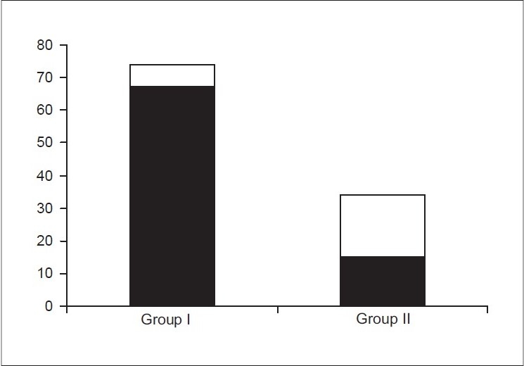
Among a total of 116 patients in the study group; group I had 78 patients, 7 (9%) required surgery; group II had 38 patients, 21(55%) required surgery. The difference in outcome between the groups was statistically significant (P = 0.002). Among those with unilateral hydronephrosis, none (0/55) with APD <15 mm required surgery, while all patients (4/4) with fetal APD> 30 mm required surgery. In those with APD between 15-30 mm, 3/19 required surgery and prolonged follow-up was required to arrive at the decision. The difference in outcome between the subgroups was statistically significant (P< 0.001, Chi-square test).
The results of our study show that simple unilateral fetal hydronephrosis runs a benign course. In the presence hydronephrosis larger than 15 mm, bilateral disease, or bladder distension, detailed postnatal evaluation and regular follow-up is warranted to plan a timely intervention. The above data could be used in prenatal counselling of these parents. Further larger studies are warranted to through more evidence.
肾积水在产前超声检查中较为常见。文献中有多种相互矛盾的预后因素,且未明确关注产后结局。本研究旨在基于产前超声评估胎儿肾积水的结局。
根据孕晚期胎儿超声检查结果,将患者分为I组(单侧肾积水)和II组(双侧肾积水、输尿管扩张、膀胱增厚等)。由一名医生按照统一方案进行产后评估和随访。比较两组间自然消退与手术干预的结局。在I组中,根据孕32周时胎儿肾盂前后径(APD)对结局进行进一步分析。
研究组共116例患者;I组78例,7例(9%)需要手术;II组38例,21例(55%)需要手术。两组间结局差异有统计学意义(P = 0.002)。在单侧肾积水患者中,APD<15 mm的无一例(0/55)需要手术,而胎儿APD>30 mm的所有患者(4/4)均需要手术。APD在15 - 30 mm之间的患者中,3/19需要手术,且需要延长随访时间才能做出决定。亚组间结局差异有统计学意义(P<0.001,卡方检验)。
我们的研究结果表明,单纯性单侧胎儿肾积水病程良性。对于肾积水大于15 mm、双侧病变或膀胱扩张的情况,有必要进行详细的产后评估和定期随访,以便及时规划干预措施。上述数据可用于这些家长的产前咨询。有必要进行进一步的大规模研究以获取更多证据。