Department of Botany, Technische Universität Darmstadt, Darmstadt, Germany.
PLoS One. 2010 Jun 15;5(6):e11112. doi: 10.1371/journal.pone.0011112.
PB1-F2 is a proapoptotic influenza A virus protein of approximately 90 amino acids in length that is located in the nucleus, cytosol and in the mitochondria membrane of infected cells. Previous studies indicated that the molecule destabilizes planar lipid bilayers and has a strong inherent tendency for multimerization. This may be correlate with its capacity to induce mitochondrial membrane depolarization.
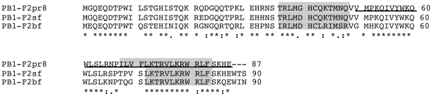
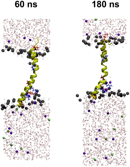
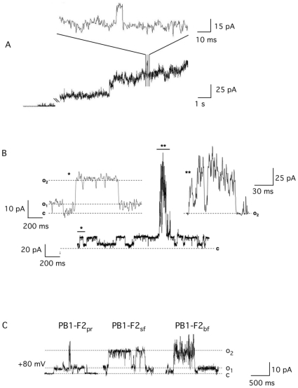
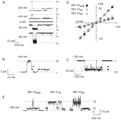
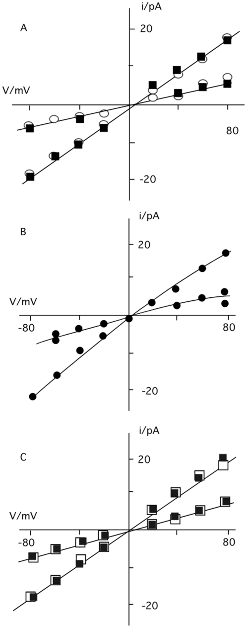
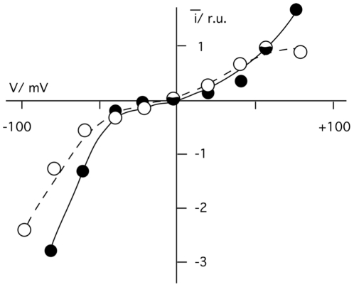
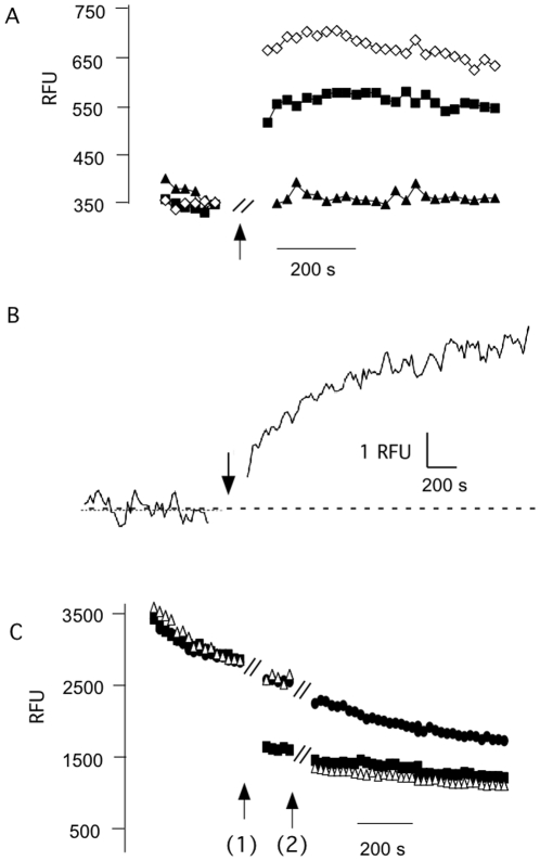
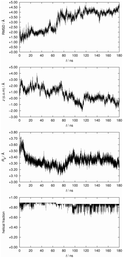
METHODOLOGY/PRINCIPAL FINDINGS: Here, we investigated whether PB1-F2 is able to form ion channels within planar lipid bilayers and microsomes. For that purpose, a set of biologically active synthetic versions of PB1-F2 (sPB1-F2) derived from the IAV isolates A/Puerto Rico/8/34(H1N1) (IAV(PR8)), from A/Brevig Mission/1/1918(H1N1) (IAV(SF2)) or the H5N1 consensus sequence (IAV(BF2)) were used. Electrical and fluorimetric measurements show that all three peptides generate in planar lipid bilayers or in liposomes, respectively, a barely selective conductance that is associated with stochastic channel type fluctuations between a closed state and at least two defined open states. Unitary channel fluctuations were also generated when a truncated protein comprising only the 37 c-terminal amino acids of sPB1-F2 was reconstituted in bilayers. Experiments were complemented by extensive molecular dynamics simulations of the truncated fragment in a lipid bilayer. The results indicate that the c-terminal region exhibits a slightly bent helical fold, which is stable and remains embedded in the bilayer for over 180 ns.
CONCLUSION/SIGNIFICANCE: The data support the idea that PB1-F2 is able to form protein channel pores with no appreciable selectivity in membranes and that the c-terminus is important for this function. This information could be important for drug development.
PB1-F2 是一种约 90 个氨基酸长的促凋亡流感 A 病毒蛋白,位于感染细胞的核、细胞质和线粒体膜中。先前的研究表明,该分子会破坏平面脂质双层的稳定性,并且具有强烈的多聚化固有倾向。这可能与其诱导线粒体膜去极化的能力有关。
方法/主要发现:在这里,我们研究了 PB1-F2 是否能够在平面脂质双层和微粒体中形成离子通道。为此,使用了一组源自 IAV 分离株 A/Puerto Rico/8/34(H1N1)(IAV(PR8))、A/Brevig Mission/1/1918(H1N1)(IAV(SF2))或 H5N1 共识序列(IAV(BF2))的具有生物活性的合成 PB1-F2 版本(sPB1-F2)。电和荧光测量表明,这三种肽在平面脂质双层或脂质体中分别产生几乎无选择性的电导,该电导与从闭合状态到至少两个定义的开放状态的随机通道类型波动相关。当在双层中重新构建仅包含 sPB1-F2 的 37 个 C 末端氨基酸的截短蛋白时,也会产生单位通道波动。实验通过对脂质双层中截短片段的广泛分子动力学模拟进行了补充。结果表明,C 末端区域表现出略微弯曲的螺旋折叠,该折叠稳定并在双层中嵌入超过 180 ns。
结论/意义:这些数据支持 PB1-F2 能够在膜中形成几乎没有选择性的蛋白质通道孔的观点,并且 C 末端对于该功能很重要。这些信息对于药物开发可能很重要。