• 文献检索
  • 文档翻译
  • 深度研究
  • 学术资讯
  • Suppr Zotero 插件Zotero 插件
  • 邀请有礼
  • 套餐&价格
  • 历史记录
应用&插件
Suppr Zotero 插件Zotero 插件浏览器插件Mac 客户端Windows 客户端微信小程序
定价
高级版会员购买积分包购买API积分包
服务
文献检索文档翻译深度研究API 文档MCP 服务
关于我们
关于 Suppr公司介绍联系我们用户协议隐私条款
关注我们

Suppr 超能文献

核心技术专利:CN118964589B侵权必究
粤ICP备2023148730 号-1Suppr @ 2026

文献检索

告别复杂PubMed语法,用中文像聊天一样搜索,搜遍4000万医学文献。AI智能推荐,让科研检索更轻松。

立即免费搜索

文件翻译

保留排版,准确专业,支持PDF/Word/PPT等文件格式,支持 12+语言互译。

免费翻译文档

深度研究

AI帮你快速写综述,25分钟生成高质量综述,智能提取关键信息,辅助科研写作。

立即免费体验

脊索细胞条件培养基刺激间充质干细胞向年轻的髓核样表型分化。

Notochordal cell conditioned medium stimulates mesenchymal stem cell differentiation toward a young nucleus pulposus phenotype.

机构信息

Cartilage Biology and Orthopaedics Branch, National Institute of Arthritis, Musculoskeletal and Skin Diseases, National Institutes of Health, Department of Health and Human Services, 50 South Drive, Bethesda, MD 20892, USA.

出版信息

Stem Cell Res Ther. 2010 Jun 16;1(2):18. doi: 10.1186/scrt18.

DOI:10.1186/scrt18
PMID:20565707
原文链接:https://pmc.ncbi.nlm.nih.gov/articles/PMC2905094/
Abstract

INTRODUCTION

Mesenchymal stem cells (MSCs) offer promise for intervertebral disc (IVD) repair and regeneration because they are easily isolated and expanded, and can differentiate into several mesenchymal tissues. Notochordal (NC) cells contribute to IVD development, incorporate into the nucleus pulposus (NP), and stimulate mature disc cells. However, there have been no studies investigating the effects of NC cells on adult stem cell differentiation. The premise of this study is that IVD regeneration is more similar to IVD development than to IVD maintenance, and we hypothesize that soluble factors from NC cells differentiate MSCs to a phenotype characteristic of nucleus pulposus (NP) cells during development. The eventual clinical goal would be to isolate or chemically/recombinantly produce the active agent to induce the therapeutic effects, and to use it as either an injectable therapy for early intervention on disc disease, or in developing appropriately pre-differentiated MSC cells in a tissue engineered NP construct.

METHODS

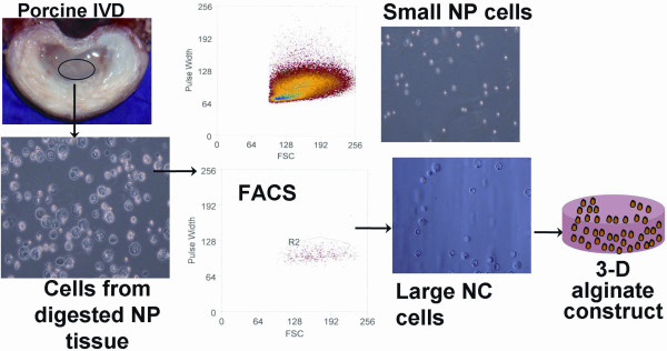
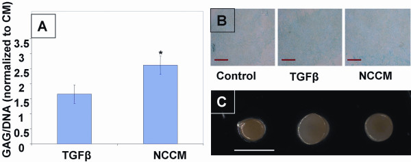
Human MSCs from bone marrow were expanded and pelleted to form high-density cultures. MSC pellets were exposed to either control medium (CM), chondrogenic medium (CM with dexamethasone and transforming growth factor, (TGF)-beta3) or notochordal cell conditioned medium (NCCM). NCCM was prepared from NC cells maintained in serum free medium for four days. After seven days culture, MSC pellets were analyzed for appearance, biochemical composition (glycosaminoglycans and DNA), and gene expression profile (sox-9, collagen types-II and III, laminin-beta1 and TIMP1(tissue inhibitor of metalloproteinases-1)).

RESULTS

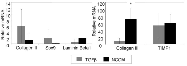
Significantly higher glycosaminoglycan accumulation was seen in NCCM treated pellets than in CM or TGFbeta groups. With NCCM treatment, increased gene expression of collagen III, and a trend of increasing expression of laminin-beta1 and decreased expression of sox-9 and collagen II relative to the TGFbeta group was observed.

CONCLUSIONS

Together, results suggest NCCM stimulates mesenchymal stem cell differentiation toward a potentially NP-like phenotype with some characteristics of the developing IVD.

摘要

简介

间充质干细胞(MSCs)为椎间盘(IVD)修复和再生提供了希望,因为它们易于分离和扩增,并能分化为几种间充质组织。脊索细胞(NC)有助于椎间盘的发育,并入髓核(NP),并刺激成熟的椎间盘细胞。然而,目前还没有研究探讨 NC 细胞对成体干细胞分化的影响。本研究的前提是,IVD 的再生更类似于 IVD 的发育,而不是 IVD 的维持,我们假设 NC 细胞的可溶性因子在发育过程中使 MSCs 分化为具有 NP 细胞表型的特征。最终的临床目标将是分离或化学/重组产生活性物质来诱导治疗效果,并将其用作早期干预椎间盘疾病的注射治疗,或在组织工程 NP 构建体中预先分化 MSC 细胞。

方法

从骨髓中扩增和沉淀人 MSC 以形成高密度培养物。MSC 沉淀暴露于对照培养基(CM)、软骨形成培养基(含地塞米松和转化生长因子-β3 的 CM)或脊索细胞条件培养基(NCCM)中。NCCM 是从在无血清培养基中维持 4 天的 NC 细胞中制备的。培养 7 天后,分析 MSC 沉淀的外观、生化成分(糖胺聚糖和 DNA)和基因表达谱(Sox-9、胶原 II 型和 III 型、层粘连蛋白-β1 和 TIMP1(金属蛋白酶抑制剂-1))。

结果

与 CM 或 TGFβ 组相比,NCCM 处理的沉淀中糖胺聚糖的积累明显更高。用 NCCM 处理后,观察到胶原 III 的基因表达增加,与 TGFβ 组相比,层粘连蛋白-β1 的表达增加趋势和 Sox-9 和胶原 II 的表达减少。

结论

综上所述,结果表明 NCCM 刺激间充质干细胞向具有潜在 NP 样表型的分化,具有一些发育中 IVD 的特征。

https://cdn.ncbi.nlm.nih.gov/pmc/blobs/977b/2905094/6d9ed2d1a7fc/scrt18-4.jpg
https://cdn.ncbi.nlm.nih.gov/pmc/blobs/977b/2905094/47460b202950/scrt18-1.jpg
https://cdn.ncbi.nlm.nih.gov/pmc/blobs/977b/2905094/dae028e84128/scrt18-2.jpg
https://cdn.ncbi.nlm.nih.gov/pmc/blobs/977b/2905094/0af0e4112b80/scrt18-3.jpg
https://cdn.ncbi.nlm.nih.gov/pmc/blobs/977b/2905094/6d9ed2d1a7fc/scrt18-4.jpg

相似文献

1
Notochordal cell conditioned medium stimulates mesenchymal stem cell differentiation toward a young nucleus pulposus phenotype.脊索细胞条件培养基刺激间充质干细胞向年轻的髓核样表型分化。
Stem Cell Res Ther. 2010 Jun 16;1(2):18. doi: 10.1186/scrt18.
2
Notochordal conditioned media from tissue increases proteoglycan accumulation and promotes a healthy nucleus pulposus phenotype in human mesenchymal stem cells.组织来源的脊索细胞条件培养液增加蛋白聚糖积累,并促进人骨髓间充质干细胞向健康的髓核样表型分化。
Arthritis Res Ther. 2011 May 31;13(3):R81. doi: 10.1186/ar3344.
3
Notochordal cells protect nucleus pulposus cells from degradation and apoptosis: implications for the mechanisms of intervertebral disc degeneration.脊索细胞保护髓核细胞免受降解和凋亡:对椎间盘退变机制的启示。
Arthritis Res Ther. 2011;13(6):R215. doi: 10.1186/ar3548. Epub 2011 Dec 29.
4
Conditioned medium derived from notochordal cell-rich nucleus pulposus tissue stimulates matrix production by canine nucleus pulposus cells and bone marrow-derived stromal cells.源自富含脊索细胞的髓核组织的条件培养基可刺激犬髓核细胞和骨髓源性基质细胞产生基质。
Tissue Eng Part A. 2015 Mar;21(5-6):1077-84. doi: 10.1089/ten.TEA.2014.0309. Epub 2014 Dec 17.
5
Activation of intervertebral disc cells by co-culture with notochordal cells, conditioned medium and hypoxia.通过与脊索细胞共培养、条件培养基和缺氧激活椎间盘细胞。
BMC Musculoskelet Disord. 2014 Dec 11;15:422. doi: 10.1186/1471-2474-15-422.
6
Intact glycosaminoglycans from intervertebral disc-derived notochordal cell-conditioned media inhibit neurite growth while maintaining neuronal cell viability.来自椎间盘源脊索细胞条件培养基的完整糖胺聚糖可抑制神经突生长,同时维持神经元细胞活力。
Spine J. 2015 May 1;15(5):1060-9. doi: 10.1016/j.spinee.2015.02.003. Epub 2015 Feb 7.
7
Human umbilical cord mesenchymal stromal cells exhibit immature nucleus pulposus cell phenotype in a laminin-rich pseudo-three-dimensional culture system.人脐带间质基质细胞在富含层粘连蛋白的拟三维培养系统中表现出未成熟的髓核细胞表型。
Stem Cell Res Ther. 2013 Oct 2;4(5):120. doi: 10.1186/scrt331.
8
Regenerative potential of TGFβ3 + Dex and notochordal cell conditioned media on degenerated human intervertebral disc cells.TGFβ3 + Dex 和脊索细胞条件培养基对退变人椎间盘细胞的再生潜能。
J Orthop Res. 2012 Mar;30(3):482-8. doi: 10.1002/jor.21534. Epub 2011 Aug 22.
9
Human iPSCs can be differentiated into notochordal cells that reduce intervertebral disc degeneration in a porcine model.人诱导多能干细胞可分化为脊索细胞,可减少猪模型中的椎间盘退变。
Theranostics. 2019 Oct 12;9(25):7506-7524. doi: 10.7150/thno.34898. eCollection 2019.
10
TGF-β3 and IGF-1 synergy ameliorates nucleus pulposus mesenchymal stem cell differentiation towards the nucleus pulposus cell type through MAPK/ERK signaling.转化生长因子-β3(TGF-β3)与胰岛素样生长因子-1(IGF-1)协同作用,通过丝裂原活化蛋白激酶/细胞外信号调节激酶(MAPK/ERK)信号通路改善髓核间充质干细胞向髓核细胞类型的分化。
Growth Factors. 2015;33(5-6):326-36. doi: 10.3109/08977194.2015.1088532. Epub 2015 Oct 2.

引用本文的文献

1
The dual effects of iron deficiency on intervertebral disc development and on acute injury-induced disc degeneration.缺铁对椎间盘发育及急性损伤诱导的椎间盘退变的双重影响。
Sci Rep. 2025 Jun 2;15(1):19323. doi: 10.1038/s41598-025-04388-4.
2
Notochordal cells: A potential therapeutic option for intervertebral disc degeneration.脊索细胞:椎间盘退变的一种潜在治疗选择。
Cell Prolif. 2024 Feb;57(2):e13541. doi: 10.1111/cpr.13541. Epub 2023 Sep 11.
3
A Review: Methodologies to Promote the Differentiation of Mesenchymal Stem Cells for the Regeneration of Intervertebral Disc Cells Following Intervertebral Disc Degeneration.

本文引用的文献

1
Morphologic complexity of the pericellular matrix in the annulus of the human intervertebral disc.人类椎间盘纤维环中细胞周围基质的形态复杂性
Biotech Histochem. 2007 Aug;82(4-5):217-25. doi: 10.1080/10520290701713999.
2
A phenotypic comparison of intervertebral disc and articular cartilage cells in the rat.大鼠椎间盘与关节软骨细胞的表型比较
Eur Spine J. 2007 Dec;16(12):2174-85. doi: 10.1007/s00586-007-0475-y. Epub 2007 Sep 5.
3
Matrix synthesis and degradation in human intervertebral disc degeneration.人类椎间盘退变中的基质合成与降解
综述:促进间充质干细胞向椎间盘退变后椎间盘细胞分化的方法。
Cells. 2023 Aug 28;12(17):2161. doi: 10.3390/cells12172161.
4
An insight on the -glycome of notochordal cell-rich porcine nucleus pulposus during maturation.对富含脊索细胞的猪髓核成熟过程中糖基化组的见解。
FASEB Bioadv. 2023 Jun 8;5(8):321-335. doi: 10.1096/fba.2023-00011. eCollection 2023 Aug.
5
An update on animal models of intervertebral disc degeneration and low back pain: Exploring the potential of artificial intelligence to improve research analysis and development of prospective therapeutics.椎间盘退变和腰痛动物模型的最新进展:探索人工智能在改善前瞻性治疗研究分析与开发方面的潜力。
JOR Spine. 2023 Jan 30;6(1):e1230. doi: 10.1002/jsp2.1230. eCollection 2023 Mar.
6
Spatiotemporal Characterization of Human Early Intervertebral Disc Formation at Single-Cell Resolution.单细胞分辨率下人早期椎间盘形成的时空特征。
Adv Sci (Weinh). 2023 May;10(14):e2206296. doi: 10.1002/advs.202206296. Epub 2023 Mar 25.
7
Two- and three-dimensional in vitro nucleus pulposus cultures: An in silico analysis of local nutrient microenvironments.二维和三维体外髓核培养:局部营养微环境的计算机模拟分析
JOR Spine. 2022 Aug 30;5(3):e1222. doi: 10.1002/jsp2.1222. eCollection 2022 Sep.
8
An esterase-responsive ibuprofen nano-micelle pre-modified embryo derived nucleus pulposus progenitor cells promote the regeneration of intervertebral disc degeneration.一种酯酶响应性布洛芬纳米胶束预修饰的胚胎源性髓核祖细胞促进椎间盘退变的再生。
Bioact Mater. 2022 Aug 14;21:69-85. doi: 10.1016/j.bioactmat.2022.07.024. eCollection 2023 Mar.
9
Encapsulation of Human-Bone-Marrow-Derived Mesenchymal Stem Cells in Small Alginate Beads Using One-Step Emulsification by Internal Gelation: In Vitro, and In Vivo Evaluation in Degenerate Intervertebral Disc Model.通过内部凝胶化一步乳化法将人骨髓间充质干细胞包裹于小海藻酸钠珠粒中:在退变椎间盘模型中的体外和体内评估
Pharmaceutics. 2022 May 31;14(6):1179. doi: 10.3390/pharmaceutics14061179.
10
Notochordal Cell-Based Treatment Strategies and Their Potential in Intervertebral Disc Regeneration.基于脊索细胞的治疗策略及其在椎间盘再生中的潜力。
Front Cell Dev Biol. 2022 Mar 14;9:780749. doi: 10.3389/fcell.2021.780749. eCollection 2021.
Biochem Soc Trans. 2007 Aug;35(Pt 4):652-5. doi: 10.1042/BST0350652.
4
Functional integrin subunits regulating cell-matrix interactions in the intervertebral disc.调节椎间盘细胞与基质相互作用的功能性整合素亚基。
J Orthop Res. 2007 Jun;25(6):829-40. doi: 10.1002/jor.20343.
5
Nucleus pulposus notochord cells secrete connective tissue growth factor and up-regulate proteoglycan expression by intervertebral disc chondrocytes.髓核脊索细胞分泌结缔组织生长因子并上调椎间盘软骨细胞的蛋白聚糖表达。
Arthritis Rheum. 2006 Dec;54(12):3859-67. doi: 10.1002/art.22258.
6
Senescence in human intervertebral discs.人类椎间盘的衰老
Eur Spine J. 2006 Aug;15 Suppl 3(Suppl 3):S312-6. doi: 10.1007/s00586-006-0126-8. Epub 2006 Jun 14.
7
Notochord cells regulate intervertebral disc chondrocyte proteoglycan production and cell proliferation.脊索细胞调节椎间盘软骨细胞蛋白聚糖的产生和细胞增殖。
Spine (Phila Pa 1976). 2006 May 1;31(10):1094-9. doi: 10.1097/01.brs.0000216593.97157.dd.
8
Notochordal cell produce and assemble extracellular matrix in a distinct manner, which may be responsible for the maintenance of healthy nucleus pulposus.脊索细胞以独特的方式产生和组装细胞外基质,这可能负责维持健康的髓核。
Spine (Phila Pa 1976). 2006 Apr 15;31(8):873-82; discussion 883. doi: 10.1097/01.brs.0000209302.00820.fd.
9
Molecular phenotypes of notochordal cells purified from immature nucleus pulposus.从未成熟髓核中纯化的脊索细胞的分子表型。
Eur Spine J. 2006 Aug;15 Suppl 3(Suppl 3):S303-11. doi: 10.1007/s00586-006-0088-x. Epub 2006 Mar 18.
10
Cytomorphology of notochordal and chondrocytic cells from the nucleus pulposus: a species comparison.来自髓核的脊索细胞和软骨细胞的细胞形态学:物种比较
J Anat. 2004 Nov;205(5):357-62. doi: 10.1111/j.0021-8782.2004.00352.x.