Department of Pathology, Pittsburgh VAMC and University of Pittsburgh, Pittsburgh, PA 15213, USA.
Mol Cancer. 2010 Jul 7;9:179. doi: 10.1186/1476-4598-9-179.
Epithelial to mesenchymal transition (EMT), implicated as a mechanism for tumor dissemination, is marked by loss of E-cadherin, disruption of cell adhesion, and induction of cell motility and invasion. In most intraductal breast carcinomas E-cadherin is regulated epigenetically via methylation of the promoter. E-cadherin expression is therefore dynamic and open to modulation by the microenvironment. In addition, it has been observed that metastatic foci commonly appear more differentiated than the primary tumor, suggesting that cancer cells may further undergo a mesenchymal to epithelial reverting transition (MErT) in the secondary organ environment following the EMT that allows for escape.
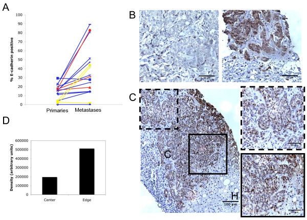
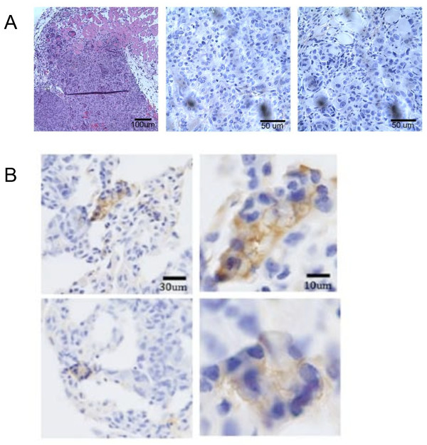
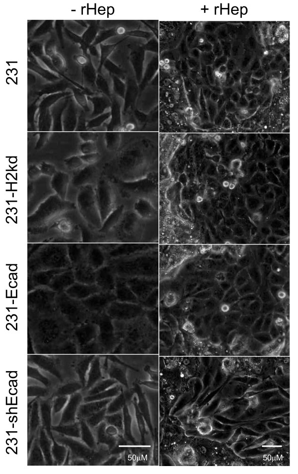
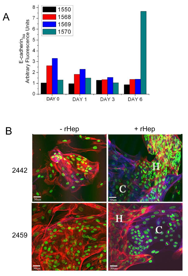
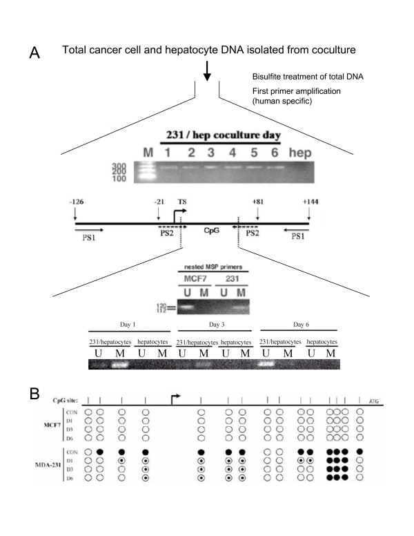
We first examined E-cadherin expression in primary breast tumors and their corresponding metastases to liver, lung and brain and discovered that 62% (10/16) of cases showed increased E-cadherin expression in the metastases compared to the primaries. These observations led to the question of whether the positive metastatic foci arose from expansion of E-cadherin-positive cells or from MErT of originally E-cadherin-negative disseminated cells. Thus, we aimed to determine whether it was possible for the mesenchymal-like MDA-MB-231 breast cancer cells to undergo an MErT through the re-expression of E-cadherin, either through exogenous introduction or induction by the microenvironment. Ectopic expression of full-length E-cadherin in MDA-MB-231 cells resulted in a morphological and functional reversion of the epithelial phenotype, with even just the cytosolic domain of E-cadherin yielding a partial phenotype. Introduction of MDA-MB-231 cells or primary explants into a secondary organ environment simulated by a hepatocyte coculture system induced E-cadherin re-expression through passive loss of methylation of the promoter. Furthermore, detection of E-cadherin-positive metastatic foci following the spontaneous metastasis of MDA-MB-231 cells injected into the mammary fat pad of mice suggests that this re-expression is functional.
Our clinical observations and experimental data indicate that the secondary organ microenvironment can induce the re-expression of E-cadherin and consequently MErT. This phenotypic change is reflected in altered cell behavior and thus may be a critical step in cell survival at metastatic sites.
上皮-间充质转化(EMT)被认为是肿瘤扩散的一种机制,其特征是 E-钙黏蛋白丢失、细胞黏附破坏以及诱导细胞运动和侵袭。在大多数导管内乳腺癌中,E-钙黏蛋白通过启动子的甲基化被表观遗传调控。因此,E-钙黏蛋白的表达是动态的,可以受到微环境的调节。此外,人们观察到转移灶通常比原发性肿瘤分化程度更高,这表明在 EMT 允许逃逸之后,癌细胞可能在次级器官环境中进一步经历间充质到上皮的逆转转化(MErT)。
我们首先检查了原发性乳腺癌及其相应的肝、肺和脑转移灶中 E-钙黏蛋白的表达,发现 62%(10/16)的病例在转移灶中 E-钙黏蛋白的表达高于原发性肿瘤。这些观察结果引发了一个问题,即阳性转移灶是由 E-钙黏蛋白阳性细胞的扩增还是由最初 E-钙黏蛋白阴性的播散细胞的 MErT 引起的。因此,我们旨在确定间充质样 MDA-MB-231 乳腺癌细胞是否有可能通过重新表达 E-钙黏蛋白(通过外源性引入或微环境诱导)来经历 MErT。在 MDA-MB-231 细胞中异位表达全长 E-钙黏蛋白导致上皮表型的形态和功能逆转,即使只是 E-钙黏蛋白的胞质结构域也产生部分表型。将 MDA-MB-231 细胞或原代外植体引入肝细胞共培养系统模拟的次级器官环境中,通过启动子的被动去甲基化诱导 E-钙黏蛋白的重新表达。此外,在将 MDA-MB-231 细胞注射到小鼠乳腺脂肪垫后自发转移的情况下检测到 E-钙黏蛋白阳性转移灶,表明这种重新表达是功能性的。
我们的临床观察和实验数据表明,次级器官微环境可以诱导 E-钙黏蛋白的重新表达,进而诱导 MErT。这种表型变化反映在改变的细胞行为上,因此可能是细胞在转移部位存活的关键步骤。