Biomedizinische NMR Forschungs GmbH am Max-Planck-Institut für biophysikalische Chemie, 37070 Göttingen, Germany.
J Cardiovasc Magn Reson. 2010 Jul 8;12(1):39. doi: 10.1186/1532-429X-12-39.
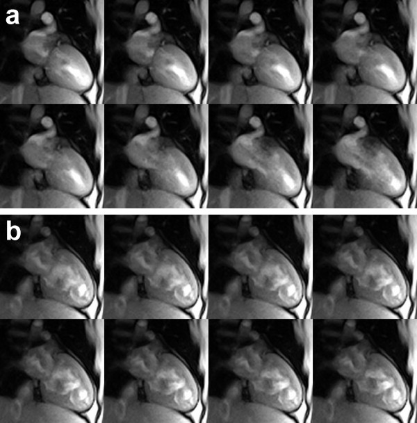
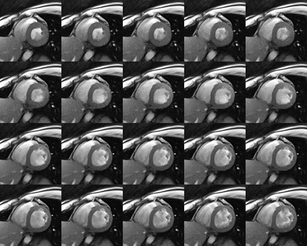
Functional assessments of the heart by dynamic cardiovascular magnetic resonance (CMR) commonly rely on (i) electrocardiographic (ECG) gating yielding pseudo real-time cine representations, (ii) balanced gradient-echo sequences referred to as steady-state free precession (SSFP), and (iii) breath holding or respiratory gating. Problems may therefore be due to the need for a robust ECG signal, the occurrence of arrhythmia and beat to beat variations, technical instabilities (e.g., SSFP "banding" artefacts), and limited patient compliance and comfort. Here we describe a new approach providing true real-time CMR with image acquisition times as short as 20 to 30 ms or rates of 30 to 50 frames per second.
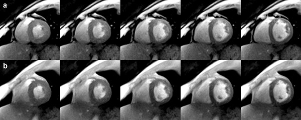
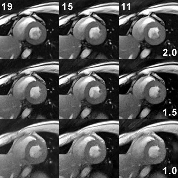
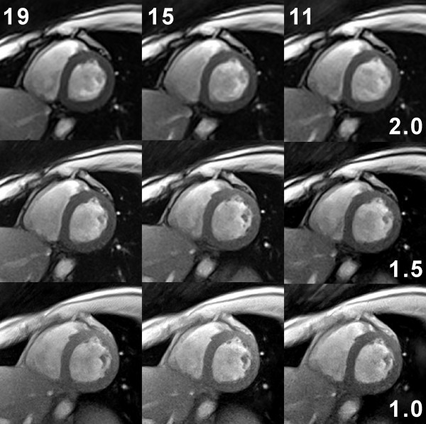
The approach relies on a previously developed real-time MR method, which combines a strongly undersampled radial FLASH CMR sequence with image reconstruction by regularized nonlinear inversion. While iterative reconstructions are currently performed offline due to limited computer speed, online monitoring during scanning is accomplished using gridding reconstructions with a sliding window at the same frame rate but with lower image quality.
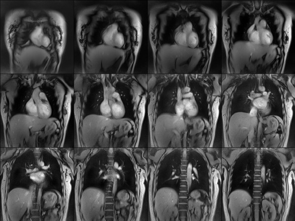
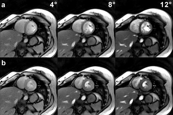
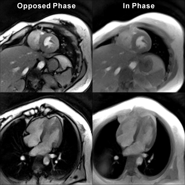
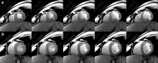
Scans of healthy young subjects were performed at 3 T without ECG gating and during free breathing. The resulting images yield T1 contrast (depending on flip angle) with an opposed-phase or in-phase condition for water and fat signals (depending on echo time). They completely avoid (i) susceptibility-induced artefacts due to the very short echo times, (ii) radiofrequency power limitations due to excitations with flip angles of 10 degrees or less, and (iii) the risk of peripheral nerve stimulation due to the use of normal gradient switching modes. For a section thickness of 8 mm, real-time images offer a spatial resolution and total acquisition time of 1.5 mm at 30 ms and 2.0 mm at 22 ms, respectively.
Though awaiting thorough clinical evaluation, this work describes a robust and flexible acquisition and reconstruction technique for real-time CMR at the ultimate limit of this technology.
动态心血管磁共振(CMR)的心脏功能评估通常依赖于(i)心电图(ECG)门控产生伪实时电影表示,(ii)平衡梯度回波序列,称为稳态自由进动(SSFP),以及(iii)屏气或呼吸门控。因此,问题可能是由于需要稳健的 ECG 信号、心律失常和心跳变化、技术不稳定性(例如,SSFP“带”伪影)以及有限的患者依从性和舒适度。在这里,我们描述了一种新方法,该方法提供了真正的实时 CMR,图像采集时间短至 20 至 30 毫秒或每秒 30 至 50 帧。
该方法依赖于以前开发的实时 MR 方法,该方法将强烈欠采样的径向 FLASH CMR 序列与正则化非线性反演的图像重建相结合。虽然由于计算机速度有限,迭代重建目前是在离线完成的,但在扫描过程中使用相同的帧率进行在线监测是通过网格重建来完成的,但图像质量较低。
在没有 ECG 门控和自由呼吸的情况下,在 3 T 对健康年轻受试者进行了扫描。所得图像产生 T1 对比度(取决于翻转角),具有水和脂肪信号的反相或同相条件(取决于回波时间)。它们完全避免了(i)由于非常短的回波时间而引起的磁化率诱导伪影,(ii)由于翻转角为 10 度或更小的激发而引起的射频功率限制,以及(iii)由于使用正常梯度切换模式而引起的外周神经刺激的风险。对于 8 毫米的切片厚度,实时图像分别在 30 毫秒和 22 毫秒时提供 1.5 毫米的空间分辨率和总采集时间,以及 2.0 毫米的空间分辨率和总采集时间。
虽然有待全面临床评估,但这项工作描述了一种用于实时 CMR 的强大且灵活的采集和重建技术,达到了该技术的极限。