Zhou Shi-you, Xie Zhao-lian, Xiao Ou, Yang Xiao-ru, Heng Boon Chin, Sato Yasufumi
Zhongshan Ophthalmic Center, Sun Yat-sen University, The State Key Laboratory of Ophthalmology, Guangzhou, China.
Mol Vis. 2010 Jul 26;16:1389-98.
To evaluate the activity of recombinant adenovirus encoding human vasohibin-1 (Ad-Vasohibin-1) on mouse corneal neovasularization induced by alkali burn.
For the treatment group, 50 mice each received subconjunctival injection (5 microl) of 10(9) plaque forming units of replication-defective Ad-Vasohibin-1. Control group mice received the same dosage of blank adenoviral vector (AdNull). Five days after injection, corneal neovascularization (CNV) was induced by placing 2.5 microl of 0.1 M NaOH on the right cornea for 30 s. Subsequently, CNV was observed and photographed every 3 days for a total duration of 9 days after the alkali burn. The percentage of neovascularized area was measured and compared with the AdNull control. The expression of human vasohibin-1 protein was detected by immunohistochemistry and western blotting at 5, 8, and 14 days after injection. The mRNA expression levels of murine vascular endothelial growth factor (Vegf), VEGF receptor 1 and 2 (Vegfr1, Vegfr2), and vasohibin-1 (Vash1) were analyzed and compared by real time quantitative reverse-transcription polymerase chain reaction.
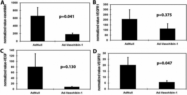
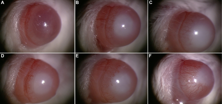
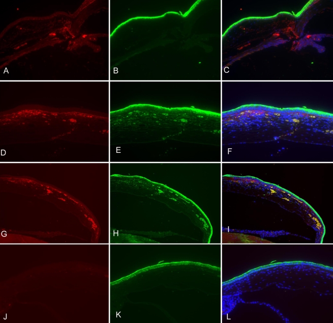
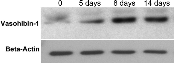
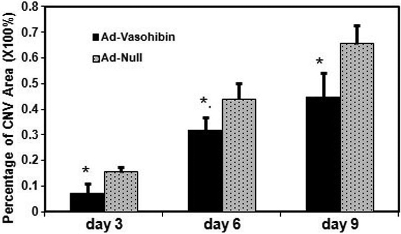
The percentage of neovascularized area within the cornea was significantly reduced in mice treated with Ad-Vasohibin-1 compared to mice treated with AdNull at every time point after alkali-induced injury (7.11%+/-3.91% and 15.48%+/-1.79% of corneal area in the treatment and control groups, respectively, on day 3; 31.64%+/-4.71% and 43.93%+/-6.15% on day 6, and 45.02%+/-9.98% and 66.24%+/-7.17% on day 9, all p<0.001). Human vasohibin-1 protein was detected at the injection sites on day 3 after corneal burn and was highly expressed in the central subepithelial stroma and co-localized with neovascularized vessels within the alkali-treated cornea on day 6. On day 9, the peripheral cornea exhibited a similar staining pattern as the central cornea, but a more intense vasohibin-1 immunostaining signal was detected in the deep stroma. Some of the vasohibin-1 stain signal diffused into the frontal and deep stroma of the central cornea and was not co-localized with new vessels. By contrast, in mice injected with AdNull or normal corneas, no vasohibin-1 stain signal was detected within the corneas. Vasohibin-1 protein expression within treated corneas was also further confirmed by western blotting on day 5. Expression appeared to peak by day 8 and was maintained at high levels until day 14. However, Vasohibin-1 protein was not detected in the corneas of normal mice or mice treated with AdNull. Real-time quantitative reverse-transcription polymerase chain reaction analysis showed that expression of Vegfr2 and endogenous Vash1 mRNA were significantly decreased in the treatment versus control group (t(1)=-2.161, p(1)=0.047; t(2)=-2.236, p(2)=0.041). In contrast, there were no significant differences in Vegf and Vegfr1 mRNA expression levels between the treatment and control groups (p>0.05 for both).
Subconjunctival injection of Ad-Vasohibin-1 significantly reduces corneal neovascularization in alkali-treated mouse corneas. This effect of anti-neovascularization may be related to the downregulation of Vegfr2 expression.
评估编码人血管抑制素-1的重组腺病毒(Ad-Vasohibin-1)对碱烧伤诱导的小鼠角膜新生血管化的作用。
治疗组50只小鼠每只接受结膜下注射(5微升)10⁹ 空斑形成单位的复制缺陷型Ad-Vasohibin-1。对照组小鼠接受相同剂量的空白腺病毒载体(AdNull)。注射5天后,通过在右眼角膜上放置2.5微升0.1 M NaOH 30秒诱导角膜新生血管化(CNV)。随后,在碱烧伤后每3天观察并拍摄CNV,共持续9天。测量新生血管化面积的百分比并与AdNull对照组进行比较。在注射后第5、8和14天通过免疫组织化学和蛋白质印迹法检测人血管抑制素-1蛋白的表达。通过实时定量逆转录聚合酶链反应分析并比较小鼠血管内皮生长因子(Vegf)、VEGF受体1和2(Vegfr1、Vegfr2)以及血管抑制素-1(Vash1)的mRNA表达水平。
与用AdNull治疗的小鼠相比,用Ad-Vasohibin-1治疗的小鼠在碱诱导损伤后的每个时间点角膜内新生血管化面积的百分比均显著降低(治疗组和对照组在第3天角膜面积分别为7.11%±3.91%和15.48%±1.79%;第6天分别为31.64%±4.71%和43.93%±6.15%;第9天分别为45.02%±9.98%和66.24%±7.17%,所有p<0.001)。角膜烧伤后第3天在注射部位检测到人血管抑制素-1蛋白,在中央上皮下基质中高表达,并且在碱处理角膜的第6天与新生血管共定位。在第9天,周边角膜呈现与中央角膜相似的染色模式,但在深层基质中检测到更强的血管抑制素-1免疫染色信号。一些血管抑制素-1染色信号扩散到中央角膜的前部和深层基质中,并且不与新血管共定位。相比之下,在注射AdNull的小鼠或正常角膜中,角膜内未检测到血管抑制素-1染色信号。在第5天通过蛋白质印迹法进一步证实了治疗角膜内血管抑制素-1蛋白的表达。表达似乎在第8天达到峰值,并在第14天之前维持在高水平。然而,在正常小鼠或用AdNull治疗的小鼠角膜中未检测到血管抑制素-1蛋白。实时定量逆转录聚合酶链反应分析表明,与对照组相比,治疗组中Vegfr2和内源性Vash1 mRNA的表达显著降低(t(1)=-2.161,p(1)=0.047;t(2)=-2.236,p(2)=0.041)。相比之下,治疗组和对照组之间Vegf和Vegfr1 mRNA表达水平没有显著差异(两者p>0.05)。
结膜下注射Ad-Vasohibin-1可显著减少碱处理小鼠角膜中的角膜新生血管化。这种抗新生血管化作用可能与Vegfr2表达的下调有关。