Thundyil John, Tang Sung-Chun, Okun Eitan, Shah Kausik, Karamyan Vardan T, Li Yu-I, Woodruff Trent M, Taylor Stephen M, Jo Dong-Gyu, Mattson Mark P, Arumugam Thiruma V
School of Biomedical Sciences, University of Queensland, Brisbane, Queensland 4072, Australia.
Exp Transl Stroke Med. 2010 Aug 11;2(1):15. doi: 10.1186/2040-7378-2-15.
Adiponectin is a hormone produced in and released from adipose cells, which has been shown to have anti-diabetic and anti-inflammatory actions in peripheral cells. Two cell surface adiponectin receptors (ADRs) mediate the majority of the known biological actions of adiponectin. Thus far, ADR expression in the brain has been demonstrated in the arcuate and the paraventricular nucleus of hypothalamus, where its activation affects food intake. Recent findings suggest that levels of circulating adiponectin increase after an ischemic stroke, but the role of adiponectin receptor activation in stroke pathogenesis and its functional outcome is unclear.
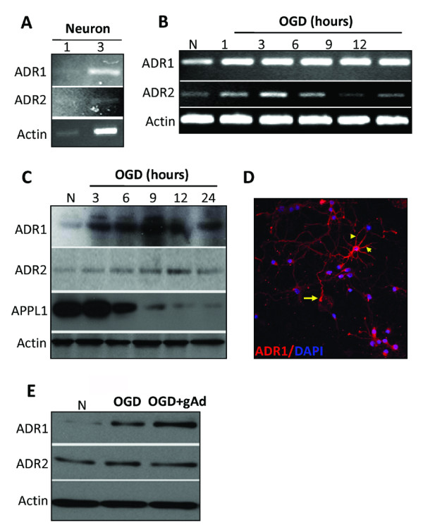
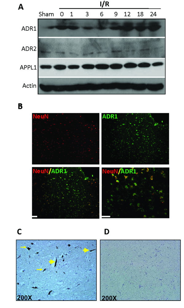
Ischemic stroke was induced in C57BL/6 mice by middle cerebral artery occlusion (MCAO) for 1 h, followed by reperfusion. Primary cortical neuronal cultures were established from individual embryonic neocortex. For glucose deprivation (GD), cultured neurons were incubated in glucose-free Locke's medium for 6, 12 or 24 h. For combined oxygen and glucose deprivation (OGD), neurons were incubated in glucose-free Locke's medium in an oxygen-free chamber with 95% N2/5% CO2 atmosphere for either 3, 6, 9, 12 or 24 h. Primary neurons and brain tissues were analysed for Adiponectin and ADRs using reverse transcriptase polymerase chain reaction (RT-PCR), immunoblot and immunochemistry methods.
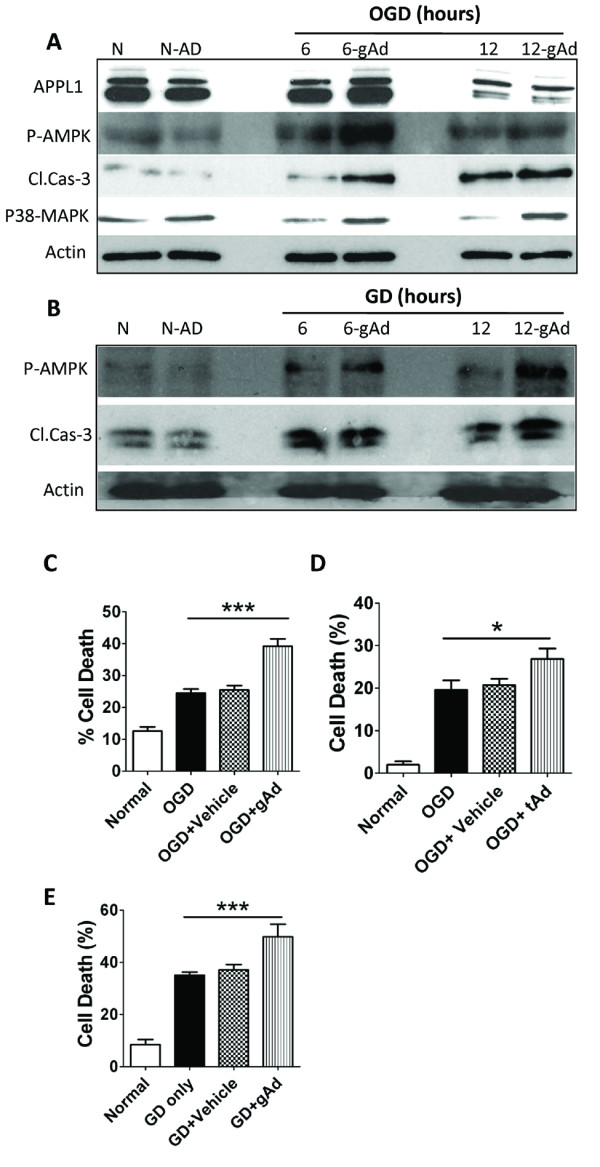
Cortical neurons express ADR1 and ADR2, and that the levels of ADR1 are increased in neurons in response to in vitro or in vivo ischemic conditions. Neurons treated with either globular or trimeric adiponectin exhibited increased vulnerability to oxygen and glucose deprivation which was associated with increased activation of a pro-apoptotic signaling cascade involving p38 mitogen-activated protein kinase (p38MAPK) and AMP-activated protein kinase (AMPK).
This study reveals a novel pathogenic role for adiponectin and adiponectin receptor activation in ischemic stroke. We show that cortical neurons express ADRs and reveal a pro-apoptotic role for ADR1 activation in neurons, which may render them vulnerable to ischemic death.
脂联素是一种由脂肪细胞产生并释放的激素,已证明其在外周细胞中具有抗糖尿病和抗炎作用。两种细胞表面脂联素受体(ADR)介导了脂联素大部分已知的生物学作用。到目前为止,已证实在下丘脑的弓状核和室旁核中有脂联素受体表达,其激活会影响食物摄入。最近的研究结果表明,缺血性中风后循环脂联素水平会升高,但脂联素受体激活在中风发病机制及其功能转归中的作用尚不清楚。
通过大脑中动脉闭塞(MCAO)1小时诱导C57BL/6小鼠发生缺血性中风,随后进行再灌注。从单个胚胎新皮质建立原代皮质神经元培养物。对于葡萄糖剥夺(GD),将培养的神经元在无葡萄糖的洛克氏培养基中孵育6、12或24小时。对于联合氧和葡萄糖剥夺(OGD),将神经元在含有95% N2/5% CO2气氛的无氧培养箱中的无葡萄糖洛克氏培养基中孵育3、6、9、12或24小时。使用逆转录聚合酶链反应(RT-PCR)、免疫印迹和免疫化学方法分析原代神经元和脑组织中的脂联素和脂联素受体。
皮质神经元表达ADR1和ADR2,并且在体外或体内缺血条件下,神经元中ADR1的水平会升高。用球状或三聚体脂联素处理的神经元对氧和葡萄糖剥夺的易感性增加,这与涉及p38丝裂原活化蛋白激酶(p38MAPK)和AMP活化蛋白激酶(AMPK)的促凋亡信号级联反应的激活增加有关。
本研究揭示了脂联素和脂联素受体激活在缺血性中风中的一种新的致病作用。我们表明皮质神经元表达脂联素受体,并揭示了ADR1激活在神经元中的促凋亡作用,这可能使它们易受缺血性死亡的影响。