Department of Neuroscience, Cell Biology, and Physiology, Boonshoft School of Medicine, Wright State University, Dayton, Ohio, USA.
J Biomed Sci. 2010 Aug 24;17 Suppl 1(Suppl 1):S10. doi: 10.1186/1423-0127-17-S1-S10.
Hippocampal slices swell and release taurine during oxidative stress. The influence of cellular signalling pathways on this process is unclear. Glutamate signalling can facilitate volume regulation in other CNS preparations. Therefore, we hypothesize activation of taurine release by oxidative stress results from tissue swelling and is coupled to activation of glutamate receptors.
Rat hippocampi were incubated at room temperature for 2 hr in artificial cerebrospinal fluid (aCSF) equilibrated with 95% O2 plus 5% CO2. For some slices, 1 mM taurine was added to the aCSF to maintain normal tissue taurine content. Slices then were perfused with aCSF at 35 degrees C and baseline data recorded before 2 mM H2O2 was added. For some studies, mannitol or inhibitors of glutamate receptors or the volume-regulated anion channel (VRAC) were added before and during H2O2 treatment. The intensity of light transmitted through the slice (the intrinsic optical signal, IOS) was determined at 1-min intervals. Samples of perfusate were collected at 2-min intervals and amino acid contents determined by HPLC. Data were analyzed by repeated measures ANOVA and post hoc Dunnett's test with significance indicated for p<0.05.
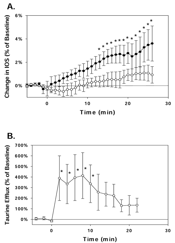
IOS of slices prepared without taurine treatment increased significantly by 3.3+/-1.3% (mean+/-SEM) during oxidative stress. Little taurine was detected in the perfusate of these slices and the rate of taurine efflux did not change during H2O2 exposure. The alpha-amino-3-hydroxyl-5-methyl-4-isoxazole-propionate antagonist, 25 microM CNQX, but not the N-methyl-D-aspartate (NMDA) receptor antagonist, 10 microM MK-801, inhibited the increase in IOS during H2O2 treatment. Taurine-treated slices exposed to H2O2 showed no change in IOS; however, taurine efflux increased by 335+/-178%. When these slices were perfused with hypertonic aCSF (350 mOsm) or exposed to the VRAC inhibitor, 20 microM DCPIB, no increase in the taurine efflux rate was observed during H2O2 exposure. Taurine-treated slices perfused with 10 microM MK-801 during H2O2 exposure showed a 4.6+/-1.9% increase in IOS but no increase in the taurine efflux rate.
Taurine efflux via VRAC is critical for volume regulation of hippocampal slices exposed to oxidative stress. This increased taurine efflux does not result from direct activation of the taurine release pathway by H2O2. NMDA receptor activation plays an important role in taurine release during oxidative stress.
在氧化应激条件下,海马切片会发生肿胀并释放牛磺酸。但细胞信号通路对此过程的影响尚不清楚。谷氨酸信号可以促进其他中枢神经系统制剂中的体积调节。因此,我们假设氧化应激导致的牛磺酸释放的激活源于组织肿胀,并与谷氨酸受体的激活相关联。
将大鼠海马在室温下于人工脑脊液(aCSF)中孵育 2 小时,该 aCSF 用 95%O2 和 5%CO2 平衡。对于一些切片,在 aCSF 中加入 1mM 牛磺酸以维持组织中的牛磺酸含量正常。然后将切片在 35°C 的 aCSF 中灌流,并在加入 2mM H2O2 之前记录基线数据。对于一些研究,在 H2O2 处理之前和期间加入甘露醇或谷氨酸受体抑制剂或体积调节阴离子通道(VRAC)抑制剂。每隔 1 分钟测定通过切片的光强度(固有光学信号,IOS)。每隔 2 分钟收集一次灌流液样品,并通过 HPLC 测定氨基酸含量。通过重复测量方差分析和事后 Dunnett 检验对数据进行分析,p<0.05 表示有统计学意义。
在氧化应激期间,未用牛磺酸处理的切片的 IOS 显著增加了 3.3+/-1.3%(平均值+/-SEM)。这些切片的灌流液中几乎没有检测到牛磺酸,并且在 H2O2 暴露期间牛磺酸的外排率没有变化。α-氨基-3-羟基-5-甲基-4-异恶唑丙酸拮抗剂 25μM CNQX,但不是 N-甲基-D-天冬氨酸(NMDA)受体拮抗剂 10μM MK-801,抑制了 H2O2 处理期间 IOS 的增加。暴露于 H2O2 的用牛磺酸处理的切片 IOS 没有变化;然而,牛磺酸外排增加了 335+/-178%。当这些切片用高渗 aCSF(350mOsm)或 VRAC 抑制剂 20μM DCPIB 灌流时,在 H2O2 暴露期间没有观察到牛磺酸外排率的增加。在 H2O2 暴露期间用 10μM MK-801 灌流的用牛磺酸处理的切片 IOS 增加了 4.6+/-1.9%,但牛磺酸外排率没有增加。
在氧化应激下,通过 VRAC 的牛磺酸外排对于海马切片的体积调节至关重要。这种增加的牛磺酸外排不是由 H2O2 直接激活牛磺酸释放途径引起的。NMDA 受体的激活在氧化应激期间的牛磺酸释放中起重要作用。