Wang Zheng, Reinach Peter S, Zhang Fan, Vellonen Kati-Sisko, Urtti Arto, Turner Helen, Wolosin J Mario
Department of Biological Sciences, SUNY State College of Optometry, New York, NY, USA.
Mol Vis. 2010 Aug 22;16:1696-704.
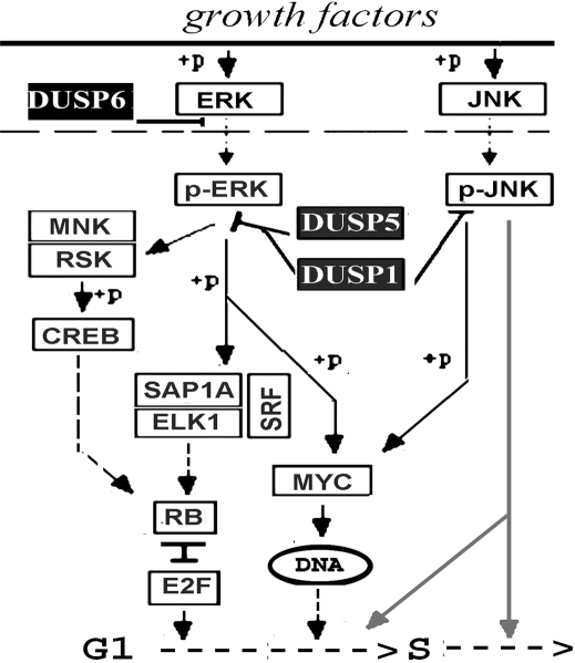
Dual specificity phosphatases (DUSPs) modulate the duration and magnitude of phospho-activation of Erk1/2, p38 and JNK1/2, the terminal kinases (TKs) of the mitogen activated protein kinase (MAPK) cascades. Three DUSPs, DUSP1, DUSP5, and DUSP6, are overexpressed in ocular surface side population stem cells (SPSCs). Our objective was to identify the impact of these enzymes on TK phosphorylation and proliferation of corneal epithelial cells.
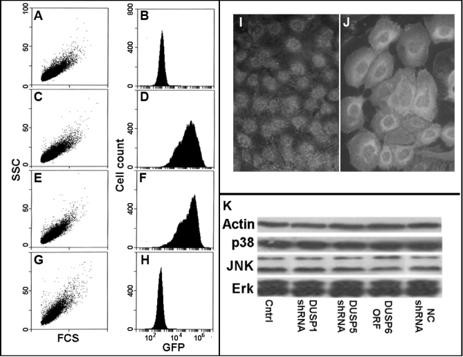
SV40 immortalized (sv) and expanded fresh human corneal epithelial cells (efHCECs) were transduced with lentivectors to elicit expression of shRNAmir against DUSP1, DUSP5, and JNK1 to thereby create the DUSP1i, DUSP5i and JNKi cell sublines, or overexpress DUSP6 (henceforth DUSP6(+)), respectively. TK phosphorylation status and proliferation rates were determined by immunoblotting and (3)H thymidine uptake.
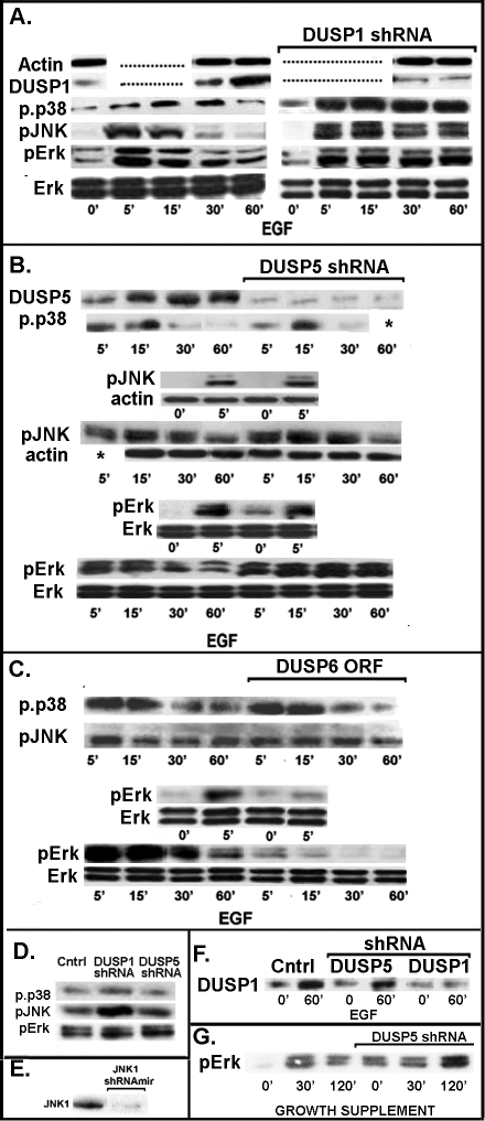
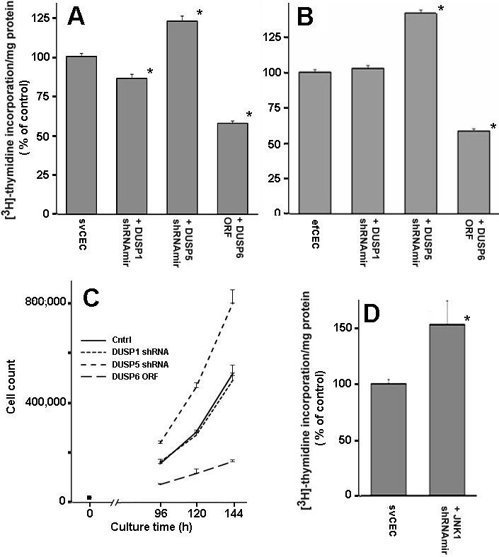
In both ef and svHCECs, EGF supplementation after a 24 h serum starvation caused a rapid 5-15 min spike in the phosphorylation of all three TK types. This was followed by gradual decreases to low phosphorylation levels within one h. These declines coincided with dramatic increases in DUSP1 and DUSP5 protein expression. In DUSP1i, the DUSP1 increase was abolished. All 3 TKs maintained high phosphorylation levels for at least 90 min and proliferation rates were unchanged from non-transduced cells. In DUSP5i, the DUSP5 protein increase was prevented, the post peak phosphorylation decrease occurred only on Erk1/2 and the proliferation rate increased by 50%-60%. In JNK1i, JNK1 was essentially knocked out and proliferation rates were also markedly elevated. At steady-state, DUSP1i maintained high levels of pJNK1/2 expression. In DUSP6(+) Erk1/2 phosphorylation was prevented and proliferation rates decreased to less than 50%.
DUSP5 and DUSP6 selectively control ERK pathway activity and proliferation. The lack of an effect of DUSP1 knockdown on proliferation can be attributed to its pan-MAPK effect. The expected augmented proliferative response due to enhanced and prolonged phosphorylation of Erk1/2 following DUSP1 knockdown does not occur because a pJNK1/2 antiproliferative effect is simultaneously unleashed.
双特异性磷酸酶(DUSPs)可调节有丝分裂原活化蛋白激酶(MAPK)级联反应的终末激酶(TKs)即Erk1/2、p38和JNK1/2磷酸化激活的持续时间和幅度。三种双特异性磷酸酶,即DUSP1、DUSP5和DUSP6,在眼表侧群干细胞(SPSCs)中过表达。我们的目的是确定这些酶对角膜上皮细胞TK磷酸化和增殖的影响。
用慢病毒载体转导猿猴病毒40(SV40)永生化(sv)并扩增的新鲜人角膜上皮细胞(efHCECs),以诱导针对DUSP1、DUSP5和JNK1的shRNAmir表达,从而分别创建DUSP1i、DUSP5i和JNKi细胞亚系,或过表达DUSP6(以下简称DUSP6(+))。通过免疫印迹和³H胸苷摄取来确定TK磷酸化状态和增殖率。
在efHCECs和svHCECs中,血清饥饿24小时后补充表皮生长因子(EGF)会导致所有三种TK类型的磷酸化在5 - 15分钟内迅速出现峰值。随后在1小时内逐渐下降至低磷酸化水平。这些下降与DUSP1和DUSP5蛋白表达的显著增加同时发生。在DUSP1i中,DUSP1的增加被消除。所有三种TK至少90分钟维持高磷酸化水平,且增殖率与未转导细胞相比无变化。在DUSP5i中,DUSP5蛋白的增加被阻止,峰值后磷酸化的下降仅发生在Erk1/2上,且增殖率提高了50% - 60%。在JNK1i中,JNK1基本被敲除,增殖率也显著升高。在稳态时,DUSP1i维持pJNK1/2的高表达水平。在DUSP6(+)中,Erk1/2的磷酸化被阻止,增殖率降至50%以下。
DUSP5和DUSP6选择性地控制ERK通路活性和增殖。DUSP1敲低对增殖无影响可归因于其泛MAPK效应。DUSP1敲低后预期的因Erk1/2磷酸化增强和延长而导致的增殖反应增强并未出现,因为同时释放了pJNK1/2的抗增殖效应。