Department of Radiation Oncology, The University of Texas MD Anderson Cancer Center, Houston, Texas, United States of America.
PLoS One. 2010 Aug 16;5(8):e12180. doi: 10.1371/journal.pone.0012180.
Normal and malignant breast tissue contains a rare population of multi-potent cells with the capacity to self-renew, referred to as stem cells, or tumor initiating cells (TIC). These cells can be enriched by growth as "mammospheres" in three-dimensional cultures.
We tested the hypothesis that human bone-marrow derived mesenchymal stem cells (MSC), which are known to support tumor growth and metastasis, increase mammosphere formation.
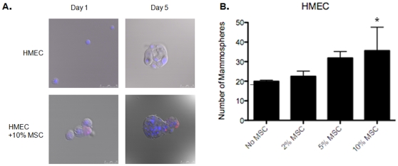
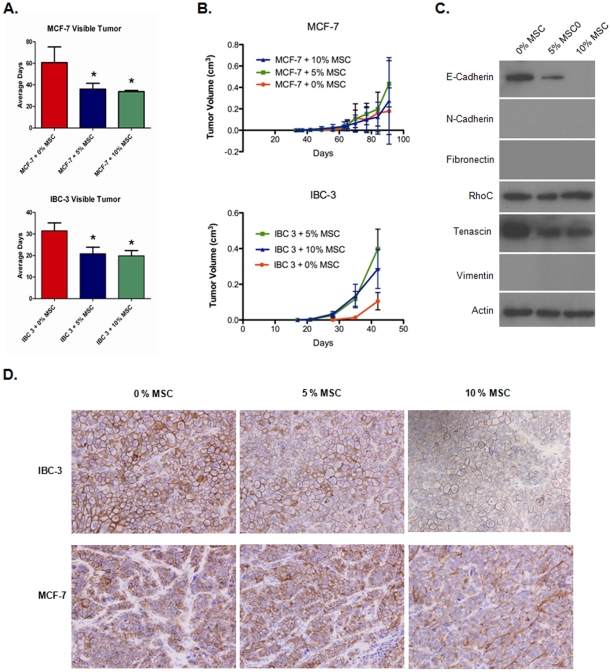
We found that MSC increased human mammary epithelial cell (HMEC) mammosphere formation in a dose-dependent manner. A similar increase in sphere formation was seen in human inflammatory (SUM149) and non-inflammatory breast cancer cell lines (MCF-7) but not in primary inflammatory breast cancer cells (MDA-IBC-3). We determined that increased mammosphere formation can be mediated by secreted factors as MSC conditioned media from MSC spheroids significantly increased HMEC, MCF-7 and SUM149 mammosphere formation by 6.4 to 21-fold. Mammospheres grown in MSC conditioned media had lower levels of the cell adhesion protein, E-cadherin, and increased expression of N-cadherin in SUM149 and HMEC cells, characteristic of a pro-invasive mesenchymal phenotype. Co-injection with MSC in vivo resulted in a reduced latency time to develop detectable MCF-7 and MDA-IBC-3 tumors and increased the growth of MDA-IBC-3 tumors. Furthermore, E-cadherin expression was decreased in MDA-IBC-3 xenografts with co-injection of MSC.
MSC increase the efficiency of primary mammosphere formation in normal and malignant breast cells and decrease E-cadherin expression, a biologic event associated with breast cancer progression and resistance to therapy.
正常和恶性乳腺组织中含有一种罕见的多能细胞群体,具有自我更新的能力,称为干细胞或肿瘤起始细胞(TIC)。这些细胞可以通过在三维培养中生长为“类乳腺球体”而被富集。
我们检验了这样一个假设,即已知能支持肿瘤生长和转移的骨髓间充质干细胞(MSC)会增加乳腺球体的形成。
我们发现,MSC 以剂量依赖的方式增加了人乳腺上皮细胞(HMEC)的乳腺球体形成。在人炎症性(SUM149)和非炎症性乳腺癌细胞系(MCF-7)中也观察到类似的球体形成增加,但在原发性炎症性乳腺癌细胞(MDA-IBC-3)中则没有。我们确定,增加的乳腺球体形成可以通过分泌因子来介导,因为 MSC 球体的条件培养基显著增加了 HMEC、MCF-7 和 SUM149 的乳腺球体形成,倍数为 6.4 到 21 倍。在 MSC 条件培养基中生长的乳腺球体具有较低水平的细胞粘附蛋白 E-钙粘蛋白,并且在 SUM149 和 HMEC 细胞中 N-钙粘蛋白的表达增加,这是一种具有侵袭前表型的特征。体内共注射 MSC 导致 MCF-7 和 MDA-IBC-3 肿瘤可检测潜伏期时间缩短,并增加 MDA-IBC-3 肿瘤的生长。此外,在与 MSC 共注射的 MDA-IBC-3 异种移植物中,E-钙粘蛋白的表达减少。
MSC 增加了正常和恶性乳腺细胞中初级乳腺球体形成的效率,并降低了 E-钙粘蛋白的表达,这是与乳腺癌进展和对治疗耐药性相关的生物学事件。