• 文献检索
  • 文档翻译
  • 深度研究
  • 学术资讯
  • Suppr Zotero 插件Zotero 插件
  • 邀请有礼
  • 套餐&价格
  • 历史记录
应用&插件
Suppr Zotero 插件Zotero 插件浏览器插件Mac 客户端Windows 客户端微信小程序
定价
高级版会员购买积分包购买API积分包
服务
文献检索文档翻译深度研究API 文档MCP 服务
关于我们
关于 Suppr公司介绍联系我们用户协议隐私条款
关注我们

Suppr 超能文献

核心技术专利:CN118964589B侵权必究
粤ICP备2023148730 号-1Suppr @ 2026

文献检索

告别复杂PubMed语法,用中文像聊天一样搜索,搜遍4000万医学文献。AI智能推荐,让科研检索更轻松。

立即免费搜索

文件翻译

保留排版,准确专业,支持PDF/Word/PPT等文件格式,支持 12+语言互译。

免费翻译文档

深度研究

AI帮你快速写综述,25分钟生成高质量综述,智能提取关键信息,辅助科研写作。

立即免费体验

PPCD1 小鼠:一种用于后多形性角膜营养不良的小鼠模型的特征描述及候选基因的鉴定。

The PPCD1 mouse: characterization of a mouse model for posterior polymorphous corneal dystrophy and identification of a candidate gene.

机构信息

McArdle Laboratory for Cancer Research, School of Medicine and Public Health, University of Wisconsin-Madison, Madison, Wisconsin, United States of America.

出版信息

PLoS One. 2010 Aug 16;5(8):e12213. doi: 10.1371/journal.pone.0012213.

DOI:10.1371/journal.pone.0012213
PMID:20808945
原文链接:https://pmc.ncbi.nlm.nih.gov/articles/PMC2922377/
Abstract

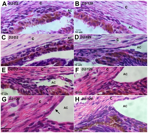
The PPCD1 mouse, a spontaneous mutant that arose in our mouse colony, is characterized by an enlarged anterior chamber resulting from metaplasia of the corneal endothelium and blockage of the iridocorneal angle by epithelialized corneal endothelial cells. The presence of stratified multilayered corneal endothelial cells with abnormal patterns of cytokeratin expression are remarkably similar to those observed in human posterior polymorphous corneal dystrophy (PPCD) and the sporadic condition, iridocorneal endothelial syndrome. Affected eyes exhibit epithelialized corneal endothelial cells, with inappropriate cytokeratin expression and proliferation over the iridocorneal angle and posterior cornea. We have termed this the "mouse PPCD1" phenotype and mapped the mouse locus for this phenotype, designated "Ppcd1", to a 6.1 Mbp interval on Chromosome 2, which is syntenic to the human Chromosome 20 PPCD1 interval. Inheritance of the mouse PPCD1 phenotype is autosomal dominant, with complete penetrance on the sensitive DBA/2J background and decreased penetrance on the C57BL/6J background. Comparative genome hybridization has identified a hemizygous 78 Kbp duplication in the mapped interval. The endpoints of the duplication are located in positions that disrupt the genes Csrp2bp and 6330439K17Rik and lead to duplication of the pseudogene LOC100043552. Quantitative reverse transcriptase-PCR indicates that expression levels of Csrp2bp and 6330439K17Rik are decreased in eyes of PPCD1 mice. Based on the observations of decreased gene expression levels, association with ZEB1-related pathways, and the report of corneal opacities in Csrp2bp(tm1a(KOMP)Wtsi) heterozygotes and embryonic lethality in nulls, we postulate that duplication of the 78 Kbp segment leading to haploinsufficiency of Csrp2bp is responsible for the mouse PPCD1 phenotype. Similarly, CSRP2BP haploinsufficiency may lead to human PPCD.

摘要

PPCD1 小鼠是我们的小鼠品系中自发出现的突变体,其特征是前房扩大,这是由于角膜内皮的化生和上皮化的角膜内皮细胞阻塞虹膜角膜角所致。具有异常细胞角蛋白表达模式的分层多层层状角膜内皮细胞与在人类后多形性角膜营养不良(PPCD)和散发性疾病、虹膜角膜内皮综合征中观察到的细胞非常相似。受影响的眼睛表现出上皮化的角膜内皮细胞,在虹膜角膜角和后角膜上表现出异常的细胞角蛋白表达和增殖。我们将这种表型称为“小鼠 PPCD1”表型,并将该表型的小鼠基因座命名为“Ppcd1”,定位于第 2 号染色体上的 6.1 Mbp 区间,与人类第 20 号染色体 PPCD1 区间是同源的。小鼠 PPCD1 表型的遗传是常染色体显性遗传,在敏感的 DBA/2J 背景下具有完全外显率,在 C57BL/6J 背景下外显率降低。比较基因组杂交鉴定出映射区间内的一个半合性 78 Kbp 重复。重复的端点位于破坏基因 Csrp2bp 和 6330439K17Rik 的位置,并导致假基因 LOC100043552 的重复。定量逆转录聚合酶链反应表明,PPCD1 小鼠眼睛中 Csrp2bp 和 6330439K17Rik 的表达水平降低。基于基因表达水平降低、与 ZEB1 相关途径的关联以及 Csrp2bp(tm1a(KOMP)Wtsi)杂合子中的角膜混浊报告和 nulls 中的胚胎致死性,我们推测导致 Csrp2bp 单倍不足的 78 Kbp 片段重复是导致小鼠 PPCD1 表型的原因。同样,CSRP2BP 单倍不足可能导致人类 PPCD。

https://cdn.ncbi.nlm.nih.gov/pmc/blobs/d622/2922377/b7fc9f9543d4/pone.0012213.g008.jpg
https://cdn.ncbi.nlm.nih.gov/pmc/blobs/d622/2922377/4ea757536aab/pone.0012213.g001.jpg
https://cdn.ncbi.nlm.nih.gov/pmc/blobs/d622/2922377/6478e4b4874c/pone.0012213.g002.jpg
https://cdn.ncbi.nlm.nih.gov/pmc/blobs/d622/2922377/36fd13513ad5/pone.0012213.g003.jpg
https://cdn.ncbi.nlm.nih.gov/pmc/blobs/d622/2922377/49de7232d2ab/pone.0012213.g004.jpg
https://cdn.ncbi.nlm.nih.gov/pmc/blobs/d622/2922377/c76b357483a7/pone.0012213.g005.jpg
https://cdn.ncbi.nlm.nih.gov/pmc/blobs/d622/2922377/3c4f818aa793/pone.0012213.g006.jpg
https://cdn.ncbi.nlm.nih.gov/pmc/blobs/d622/2922377/a2e60431e1c0/pone.0012213.g007.jpg
https://cdn.ncbi.nlm.nih.gov/pmc/blobs/d622/2922377/b7fc9f9543d4/pone.0012213.g008.jpg

相似文献

1
The PPCD1 mouse: characterization of a mouse model for posterior polymorphous corneal dystrophy and identification of a candidate gene.PPCD1 小鼠:一种用于后多形性角膜营养不良的小鼠模型的特征描述及候选基因的鉴定。
PLoS One. 2010 Aug 16;5(8):e12213. doi: 10.1371/journal.pone.0012213.
2
Association of a Chromosomal Rearrangement Event with Mouse Posterior Polymorphous Corneal Dystrophy and Alterations in Csrp2bp, Dzank1, and Ovol2 Gene Expression.染色体重排事件与小鼠后部多形性角膜营养不良及Csrp2bp、Dzank1和Ovol2基因表达改变的关联
PLoS One. 2016 Jun 16;11(6):e0157577. doi: 10.1371/journal.pone.0157577. eCollection 2016.
3
Exclusion of positional candidate gene coding region mutations in the common posterior polymorphous corneal dystrophy 1 candidate gene interval.排除常见后部多形性角膜营养不良1候选基因区间内的位置候选基因编码区突变。
Cornea. 2009 Aug;28(7):801-7. doi: 10.1097/ICO.0b013e31819672fb.
4
Genetic analysis of chromosome 20-related posterior polymorphous corneal dystrophy: genetic heterogeneity and exclusion of three candidate genes.20号染色体相关的后部多形性角膜营养不良的基因分析:基因异质性及三个候选基因的排除
Mol Vis. 2008 Jan 16;14:71-80.
5
Retinal pathology in the PPCD1 mouse.PPCD1小鼠的视网膜病理学
PLoS One. 2017 Oct 5;12(10):e0185094. doi: 10.1371/journal.pone.0185094. eCollection 2017.
6
Alterations in GRHL2-OVOL2-ZEB1 axis and aberrant activation of Wnt signaling lead to altered gene transcription in posterior polymorphous corneal dystrophy.GRHL2-OVOL2-ZEB1 轴的改变和 Wnt 信号的异常激活导致后多形性角膜营养不良中基因转录的改变。
Exp Eye Res. 2019 Nov;188:107696. doi: 10.1016/j.exer.2019.107696. Epub 2019 Jun 21.
7
Confirmation of the OVOL2 Promoter Mutation c.-307T>C in Posterior Polymorphous Corneal Dystrophy 1.后极性多形性角膜营养不良1中OVOL2启动子突变c.-307T>C的确认
PLoS One. 2017 Jan 3;12(1):e0169215. doi: 10.1371/journal.pone.0169215. eCollection 2017.
8
Zeb1 mutant mice as a model of posterior corneal dystrophy.Zeb1突变小鼠作为后角膜营养不良的模型。
Invest Ophthalmol Vis Sci. 2008 May;49(5):1843-9. doi: 10.1167/iovs.07-0789.
9
Ovol2 promoter mutations in mice and human illuminate species-specific phenotypic divergence.Ovol2 启动子突变在小鼠和人类中揭示了种间特异性表型分化。
Hum Mol Genet. 2024 Feb 28;33(6):491-500. doi: 10.1093/hmg/ddad195.
10
Ectopic GRHL2 Expression Due to Non-coding Mutations Promotes Cell State Transition and Causes Posterior Polymorphous Corneal Dystrophy 4.非编码突变导致的异位 GRHL2 表达促进细胞状态转变并导致 4 型后部多形性角膜营养不良。
Am J Hum Genet. 2018 Mar 1;102(3):447-459. doi: 10.1016/j.ajhg.2018.02.002.

引用本文的文献

1
Preclinical Models for Studying Fuchs Endothelial Corneal Dystrophy.用于研究富克斯角膜内皮营养不良的临床前模型
Cells. 2025 Mar 28;14(7):505. doi: 10.3390/cells14070505.
2
Animal models of corneal endothelial dysfunction to facilitate development of novel therapies.角膜内皮功能障碍的动物模型,以促进新型疗法的开发。
Ann Transl Med. 2021 Aug;9(15):1271. doi: 10.21037/atm-20-4389.
3
Retinal pathology in the PPCD1 mouse.PPCD1小鼠的视网膜病理学

本文引用的文献

1
Missense mutations in TCF8 cause late-onset Fuchs corneal dystrophy and interact with FCD4 on chromosome 9p.TCF8 错义突变导致迟发性 Fuchs 角膜营养不良,并与 9p 染色体上的 FCD4 相互作用。
Am J Hum Genet. 2010 Jan;86(1):45-53. doi: 10.1016/j.ajhg.2009.12.001. Epub 2009 Dec 31.
2
Phenotypic characterisation and ZEB1 mutational analysis in posterior polymorphous corneal dystrophy in a New Zealand population.新西兰人群中后部多形性角膜营养不良的表型特征及ZEB1突变分析
Mol Vis. 2009 Dec 3;15:2544-53.
3
The clinical spectrum between posterior polymorphous dystrophy and iridocorneal endothelial syndromes.
PLoS One. 2017 Oct 5;12(10):e0185094. doi: 10.1371/journal.pone.0185094. eCollection 2017.
4
Confirmation of the OVOL2 Promoter Mutation c.-307T>C in Posterior Polymorphous Corneal Dystrophy 1.后极性多形性角膜营养不良1中OVOL2启动子突变c.-307T>C的确认
PLoS One. 2017 Jan 3;12(1):e0169215. doi: 10.1371/journal.pone.0169215. eCollection 2017.
5
Association of a Chromosomal Rearrangement Event with Mouse Posterior Polymorphous Corneal Dystrophy and Alterations in Csrp2bp, Dzank1, and Ovol2 Gene Expression.染色体重排事件与小鼠后部多形性角膜营养不良及Csrp2bp、Dzank1和Ovol2基因表达改变的关联
PLoS One. 2016 Jun 16;11(6):e0157577. doi: 10.1371/journal.pone.0157577. eCollection 2016.
6
Genetics of the corneal endothelial dystrophies: an evidence-based review.角膜内皮营养不良的遗传学:基于证据的综述。
Clin Genet. 2013 Aug;84(2):109-19. doi: 10.1111/cge.12191. Epub 2013 Jun 10.
Optometry. 2009 Aug;80(8):431-6. doi: 10.1016/j.optm.2009.02.009.
4
Exclusion of positional candidate gene coding region mutations in the common posterior polymorphous corneal dystrophy 1 candidate gene interval.排除常见后部多形性角膜营养不良1候选基因区间内的位置候选基因编码区突变。
Cornea. 2009 Aug;28(7):801-7. doi: 10.1097/ICO.0b013e31819672fb.
5
The double-histone-acetyltransferase complex ATAC is essential for mammalian development.双组蛋白乙酰转移酶复合物ATAC对哺乳动物发育至关重要。
Mol Cell Biol. 2009 Mar;29(5):1176-88. doi: 10.1128/MCB.01599-08. Epub 2008 Dec 22.
6
The role of the ZEB family of transcription factors in development and disease.转录因子ZEB家族在发育和疾病中的作用。
Cell Mol Life Sci. 2009 Mar;66(5):773-87. doi: 10.1007/s00018-008-8465-8.
7
Human ATAC Is a GCN5/PCAF-containing acetylase complex with a novel NC2-like histone fold module that interacts with the TATA-binding protein.人源ATAC是一种含有GCN5/PCAF的乙酰化酶复合物,具有一个与TATA结合蛋白相互作用的新型NC2样组蛋白折叠模块。
J Biol Chem. 2008 Dec 5;283(49):33808-15. doi: 10.1074/jbc.M806936200. Epub 2008 Oct 6.
8
Zeb1 mutant mice as a model of posterior corneal dystrophy.Zeb1突变小鼠作为后角膜营养不良的模型。
Invest Ophthalmol Vis Sci. 2008 May;49(5):1843-9. doi: 10.1167/iovs.07-0789.
9
Genetic analysis of chromosome 20-related posterior polymorphous corneal dystrophy: genetic heterogeneity and exclusion of three candidate genes.20号染色体相关的后部多形性角膜营养不良的基因分析:基因异质性及三个候选基因的排除
Mol Vis. 2008 Jan 16;14:71-80.
10
Congenital corneal opacities: a review with a focus on genetics.先天性角膜混浊:聚焦遗传学的综述
Semin Ophthalmol. 2007 Oct-Dec;22(4):241-6. doi: 10.1080/08820530701745157.