Center for Biomedical Engineering, Department of Medicine, Brigham and Women's Hospital, Harvard Medical School, 65 Landsdowne Street, Cambridge, MA 02139, USA.
Biofabrication. 2010 Sep;2(3):035003. doi: 10.1088/1758-5082/2/3/035003. Epub 2010 Sep 8.
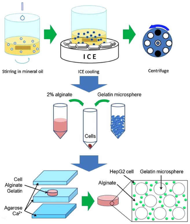
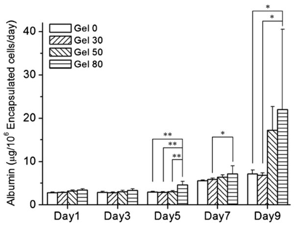
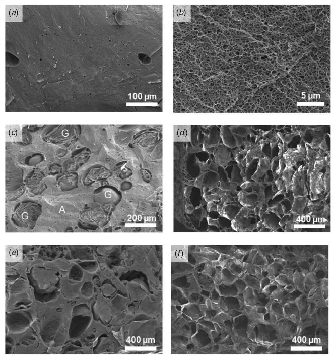
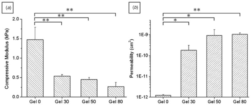
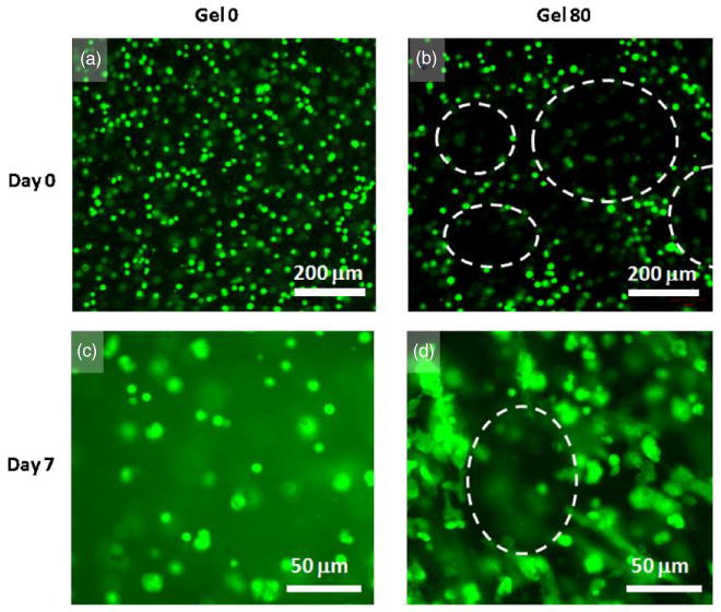
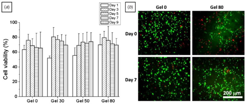
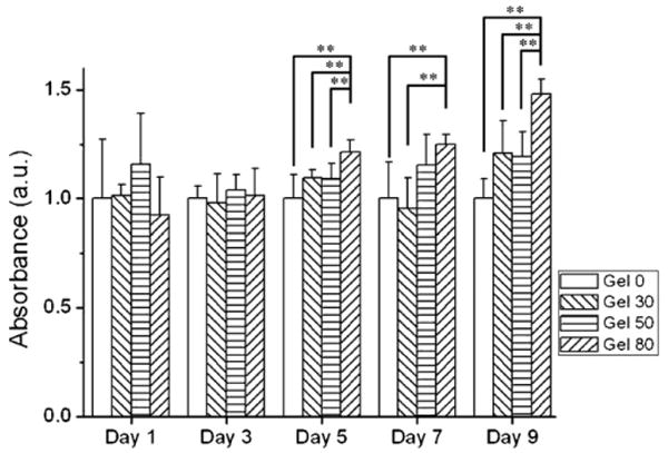
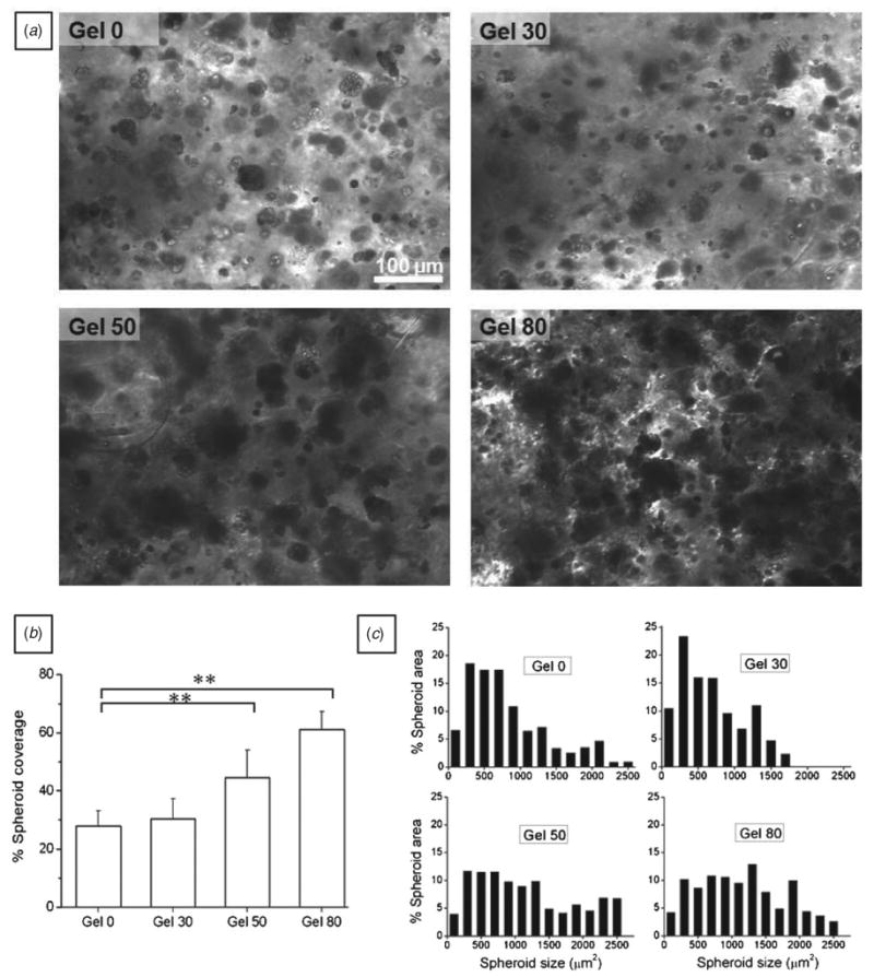
For tissue engineering applications, scaffolds should be porous to enable rapid nutrient and oxygen transfer while providing a three-dimensional (3D) microenvironment for the encapsulated cells. This dual characteristic can be achieved by fabrication of porous hydrogels that contain encapsulated cells. In this work, we developed a simple method that allows cell encapsulation and pore generation inside alginate hydrogels simultaneously. Gelatin beads of 150-300 microm diameter were used as a sacrificial porogen for generating pores within cell-laden hydrogels. Gelation of gelatin at low temperature (4 degrees C) was used to form beads without chemical crosslinking and their subsequent dissolution after cell encapsulation led to generation of pores within cell-laden hydrogels. The pore size and porosity of the scaffolds were controlled by the gelatin bead size and their volume ratio, respectively. Fabricated hydrogels were characterized for their internal microarchitecture, mechanical properties and permeability. Hydrogels exhibited a high degree of porosity with increasing gelatin bead content in contrast to nonporous alginate hydrogel. Furthermore, permeability increased by two to three orders while compressive modulus decreased with increasing porosity of the scaffolds. Application of these scaffolds for tissue engineering was tested by encapsulation of hepatocarcinoma cell line (HepG2). All the scaffolds showed similar cell viability; however, cell proliferation was enhanced under porous conditions. Furthermore, porous alginate hydrogels resulted in formation of larger spheroids and higher albumin secretion compared to nonporous conditions. These data suggest that porous alginate hydrogels may have provided a better environment for cell proliferation and albumin production. This may be due to the enhanced mass transfer of nutrients, oxygen and waste removal, which is potentially beneficial for tissue engineering and regenerative medicine applications.
对于组织工程应用,支架应为多孔的,以允许快速的营养物质和氧气转移,同时为封装的细胞提供三维(3D)微环境。通过制造包含封装细胞的多孔水凝胶可以实现这种双重特性。在这项工作中,我们开发了一种简单的方法,可以同时在藻酸盐水凝胶中进行细胞封装和孔生成。使用直径为 150-300 微米的明胶珠作为牺牲性成孔剂,在细胞负载水凝胶中生成孔。在低温(4°C)下进行明胶凝胶化,以形成无化学交联的珠体,随后在细胞封装后将其溶解,导致细胞负载水凝胶中生成孔。支架的孔径和孔隙率分别通过明胶珠的大小和其体积比来控制。对所制造的水凝胶进行了内部微观结构、力学性能和渗透性的表征。与非多孔藻酸盐水凝胶相比,随着明胶珠含量的增加,水凝胶表现出高的多孔性和高的孔隙率。此外,渗透性提高了两个到三个数量级,而压缩模量随着支架孔隙率的增加而降低。通过封装肝癌细胞系(HepG2)来测试这些支架在组织工程中的应用。所有支架均表现出相似的细胞活力;然而,在多孔条件下细胞增殖得到增强。此外,与非多孔条件相比,多孔藻酸盐水凝胶导致形成更大的球体和更高的白蛋白分泌。这些数据表明,多孔藻酸盐水凝胶可能为细胞增殖和白蛋白产生提供了更好的环境。这可能是由于营养物质、氧气和废物的传质增强,这对于组织工程和再生医学应用可能是有益的。