H. Lee Moffitt Cancer Center and Research Institute, University of South Florida, Tampa, FL 33612, USA.
J Exp Med. 2010 Oct 25;207(11):2439-53. doi: 10.1084/jem.20100587. Epub 2010 Sep 27.
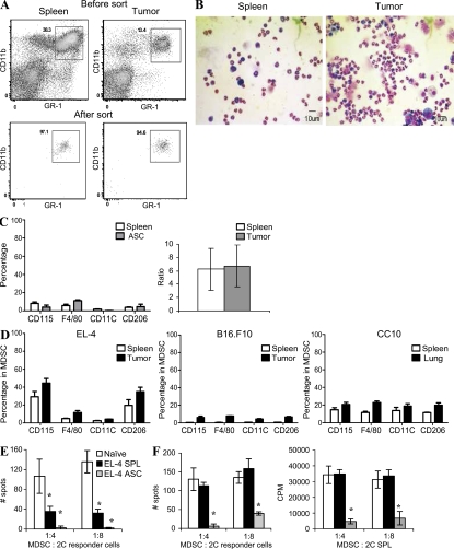
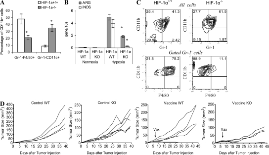
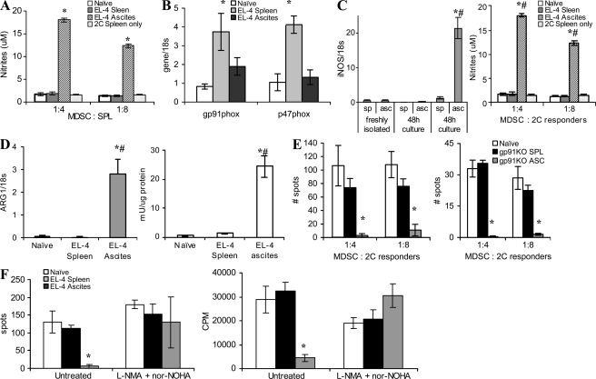
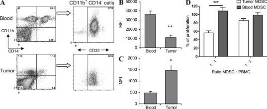
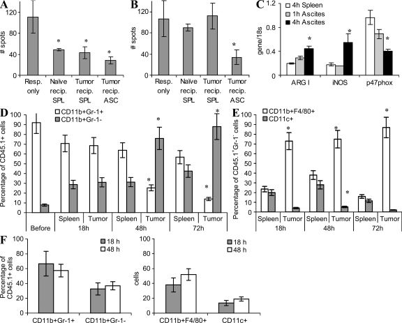
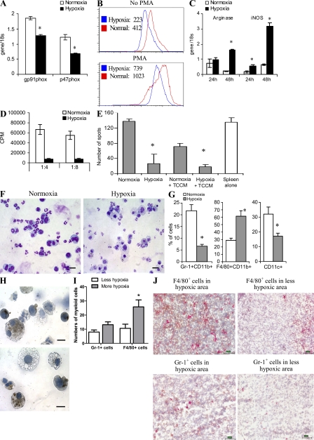
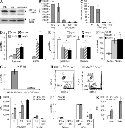
Myeloid-derived suppressor cells (MDSCs) are a major component of the immune-suppressive network described in cancer and many other pathological conditions. We demonstrate that although MDSCs from peripheral lymphoid organs and the tumor site share similar phenotype and morphology, these cells display profound functional differences. MDSC from peripheral lymphoid organs suppressed antigen-specific CD8(+) T cells but failed to inhibit nonspecific T cell function. In sharp contrast, tumor MDSC suppressed both antigen-specific and nonspecific T cell activity. The tumor microenvironment caused rapid and dramatic up-regulation of arginase I and inducible nitric oxide synthase in MDSC, which was accompanied by down-regulation of nicotinamide adenine dinucleotide phosphate-oxidase and reactive oxygen species in these cells. In contrast to MDSC from the spleen, MDSC from the tumor site rapidly differentiated into macrophages. Exposure of spleen MDSC to hypoxia resulted in the conversion of these cells to nonspecific suppressors and their preferential differentiation to macrophages. Hypoxia-inducible factor (HIF) 1α was found to be primarily responsible for the observed effects of the tumor microenvironment on MDSC differentiation and function. Thus, hypoxia via HIF-1α dramatically alters the function of MDSC in the tumor microenvironment and redirects their differentiation toward tumor-associated macrophages, hence providing a mechanistic link between different myeloid suppressive cells in the tumor microenvironment.
髓系来源的抑制细胞(MDSCs)是癌症和许多其他病理状况中描述的免疫抑制网络的主要组成部分。我们证明,尽管来自外周淋巴器官和肿瘤部位的 MDSC 具有相似的表型和形态,但这些细胞表现出明显的功能差异。外周淋巴器官的 MDSC 抑制抗原特异性 CD8(+) T 细胞,但未能抑制非特异性 T 细胞功能。与此形成鲜明对比的是,肿瘤 MDSC 抑制了抗原特异性和非特异性 T 细胞的活性。肿瘤微环境导致 MDSC 中精氨酸酶 I 和诱导型一氧化氮合酶的快速和显著上调,同时伴随着这些细胞中烟酰胺腺嘌呤二核苷酸磷酸氧化酶和活性氧的下调。与来自脾脏的 MDSC 不同,来自肿瘤部位的 MDSC 迅速分化为巨噬细胞。将脾脏 MDSC 暴露于缺氧条件下,导致这些细胞转化为非特异性抑制剂,并优先分化为巨噬细胞。缺氧诱导因子 1α(HIF-1α)被发现主要负责肿瘤微环境对 MDSC 分化和功能的观察到的影响。因此,缺氧通过 HIF-1α 显著改变了 MDSC 在肿瘤微环境中的功能,并将其分化方向转向肿瘤相关的巨噬细胞,从而为肿瘤微环境中不同髓系抑制细胞之间提供了一种机制联系。