Department of Radiology (Hs-224), Erasmus MC-University Medical Centre Rotterdam, PO Box 2040, 3000 CA, Rotterdam, the Netherlands.
Neuroradiology. 2011 Aug;53(8):553-63. doi: 10.1007/s00234-010-0774-6. Epub 2010 Oct 6.
After minor head injury (MHI), post-concussive symptoms commonly occur. The purpose of this study was to correlate the severity of post-concussive symptoms in MHI patients with MRI measures of microstructural brain injury, namely mean diffusivity (MD) and fractional anisotropy (FA), as well as the presence of microhaemorrhages.
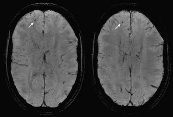
Twenty MHI patients and 12 healthy controls were scanned at 3 T using diffusion tensor imaging (DTI) and high-resolution gradient recalled echo (HRGRE) T2*-weighted sequences. One patient was excluded from the analysis because of bilateral subdural haematomas. DTI data were preprocessed using Tract Based Spatial Statistics. The resulting MD and FA images were correlated with the severity of post-concussive symptoms evaluated with the Rivermead Postconcussion Symptoms Questionnaire. The number and location of microhaemorrhages were assessed on the HRGRE T2*-weighted images.
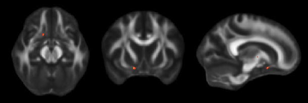
Comparing patients with controls, there were no differences in MD. FA was decreased in the right temporal subcortical white matter. MD was increased in association with the severity of post-concussive symptoms in the inferior fronto-occipital fasciculus (IFO), the inferior longitudinal fasciculus and the superior longitudinal fasciculus. FA was reduced in association with the severity of post-concussive symptoms in the uncinate fasciculus, the IFO, the internal capsule and the corpus callosum, as well as in the parietal and frontal subcortical white matter. Microhaemorrhages were observed in one patient only.
The severity of post-concussive symptoms after MHI was significantly correlated with a reduction of white matter integrity, providing evidence of microstructural brain injury as a neuropathological substrate of the post-concussion syndrome.
轻度头部外伤(MHI)后常出现脑震荡后症状。本研究旨在将 MHI 患者脑震荡后症状的严重程度与磁共振成像(MRI)测量的微结构脑损伤相关联,即平均扩散系数(MD)和各向异性分数(FA),以及微出血的存在。
20 名 MHI 患者和 12 名健康对照者在 3T 上使用弥散张量成像(DTI)和高分辨率梯度回波(HRGRE)T2*-加权序列进行扫描。由于双侧硬膜下血肿,1 名患者被排除在分析之外。使用基于束的空间统计学对 DTI 数据进行预处理。将得到的 MD 和 FA 图像与用 Rivermead 脑震荡后症状问卷评估的脑震荡后症状的严重程度进行相关性分析。在 HRGRE T2*-加权图像上评估微出血的数量和位置。
与对照组相比,患者的 MD 无差异。右侧颞叶皮质下白质的 FA 降低。MD 与脑震荡后症状的严重程度相关联,在下额枕下束(IFO)、下纵束和上纵束中增加。FA 与脑震荡后症状的严重程度相关联,在钩束、IFO、内囊和胼胝体,以及顶叶和额叶皮质下白质中减少。只有 1 名患者观察到微出血。
MHI 后脑震荡后症状的严重程度与白质完整性的降低显著相关,为脑震荡后综合征的神经病理学基础提供了微结构脑损伤的证据。