Department of Medicine, University of California San Diego, USA.
Respir Res. 2010 Nov 1;11(1):154. doi: 10.1186/1465-9921-11-154.
In this study we examined the role of Siglec-F, a receptor highly expressed on eosinophils, in contributing to mucus expression, airway remodeling, and Siglec-F ligand expression utilizing Siglec-F deficient mice exposed to chronic allergen challenge.
Wild type (WT) and Siglec-F deficient mice were sensitized and challenged chronically with OVA for one month. Levels of airway inflammation (eosinophils), Siglec-F ligand expresion and remodeling (mucus, fibrosis, smooth muscle thickness, extracellular matrix protein deposition) were assessed in lung sections by image analysis and immunohistology. Airway hyperreactivity to methacholine was assessed in intubated and ventilated mice.
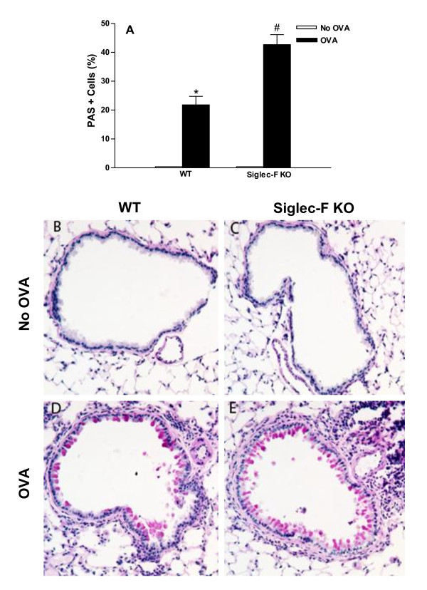
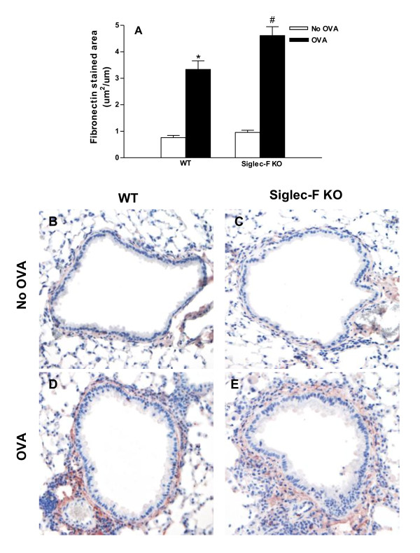
Siglec-F deficient mice challenged with OVA for one month had significantly increased numbers of BAL and peribronchial eosinophils compared to WT mice which was associated with a significant increase in mucus expression as assessed by the number of periodic acid Schiff positive airway epithelial cells. In addition, OVA challenged Siglec-F deficient mice had significantly increased levels of peribronchial fibrosis (total lung collagen, area of peribronchial trichrome staining), as well as increased numbers of peribronchial TGF-β1+ cells, and increased levels of expression of the extracellular matrix protein fibronectin compared to OVA challenged WT mice. Lung sections immunostained with a Siglec-Fc to detect Siglec-F ligand expression demonstrated higher levels of expression of the Siglec-F ligand in the peribronchial region in OVA challenged Siglec-F deficient mice compared to WT mice. WT and Siglec-F deficient mice challenged intranasally with IL-4 or IL-13 had significantly increased levels of airway epithelial Siglec-F ligand expression, whereas this was not observed in WT or Siglec-F deficient mice challenged with TNF-α. There was a significant increase in the thickness of the peribronchial smooth muscle layer in OVA challenged Siglec-F deficient mice, but this was not associated with significant increased airway hyperreactivity compared to WT mice.
Overall, this study demonstrates an important role for Siglec-F in modulating levels of chronic eosinophilic airway inflammation, peribronchial fibrosis, thickness of the smooth muscle layer, mucus expression, fibronectin, and levels of peribronchial Siglec-F ligands suggesting that Siglec-F may normally function to limit levels of chronic eosinophilic inflammation and remodeling. In addition, IL-4 and IL-13 are important regulators of Siglec-F ligand expression by airway epithelium.
在这项研究中,我们利用 Siglec-F 缺陷小鼠,研究了在慢性变应原刺激下 Siglec-F(一种在嗜酸性粒细胞上高度表达的受体)在促进黏液表达、气道重塑和 Siglec-F 配体表达中的作用。
野生型(WT)和 Siglec-F 缺陷型小鼠经致敏和慢性 OVA 攻击一个月。通过图像分析和免疫组织化学,评估肺组织切片中气道炎症(嗜酸性粒细胞)、Siglec-F 配体表达和重塑(黏液、纤维化、平滑肌厚度、细胞外基质蛋白沉积)。通过气管插管和机械通气评估气道对乙酰甲胆碱的高反应性。
与 WT 小鼠相比,经 OVA 攻击一个月的 Siglec-F 缺陷型小鼠 BAL 和支气管周围的嗜酸性粒细胞数量明显增加,这与气道上皮细胞过碘酸希夫(PAS)阳性的黏液表达明显增加有关。此外,与 OVA 攻击 WT 小鼠相比,Siglec-F 缺陷型小鼠 OVA 攻击后,支气管周围纤维化(肺胶原总量、支气管周围三染色面积)、支气管周围 TGF-β1+细胞数量以及细胞外基质蛋白纤维连接蛋白的表达水平均显著增加。用 Siglec-Fc 免疫组化染色检测 Siglec-F 配体的表达,结果显示 Siglec-F 缺陷型小鼠的支气管周围区域 Siglec-F 配体表达水平明显高于 WT 小鼠。与 TNF-α 攻击 WT 或 Siglec-F 缺陷型小鼠不同,IL-4 或 IL-13 攻击 WT 或 Siglec-F 缺陷型小鼠均显著增加气道上皮 Siglec-F 配体的表达。在 OVA 攻击 Siglec-F 缺陷型小鼠中,支气管周围平滑肌层厚度显著增加,但与 WT 小鼠相比,气道高反应性无明显增加。
总的来说,这项研究表明 Siglec-F 在调节慢性嗜酸性粒细胞性气道炎症、支气管周围纤维化、平滑肌层厚度、黏液表达、纤维连接蛋白和支气管周围 Siglec-F 配体水平方面发挥着重要作用,提示 Siglec-F 可能正常发挥作用,以限制慢性嗜酸性粒细胞炎症和重塑的程度。此外,IL-4 和 IL-13 是气道上皮 Siglec-F 配体表达的重要调节因子。