Department of Biochemistry and Department of Medicine, University of Oklahoma Health Science Center, Oklahoma City, Oklahoma, United States of America.
PLoS One. 2010 Nov 5;5(11):e15420. doi: 10.1371/journal.pone.0015420.
Redox state is a critical determinant of cell function, and any major imbalances can cause severe damage or death.
The aim of this study is to determine if AMP-activated protein kinase (AMPK), a cellular energy sensor, is activated by oxidants generated by Berberine in endothelial cells (EC).
Bovine aortic endothelial cells (BAEC) were exposed to Berberine. AMPK activity and reactive oxygen species were monitored after the incubation.
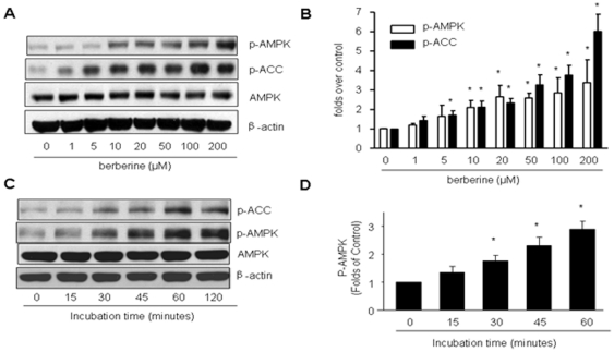
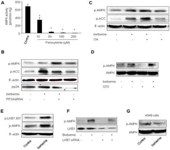
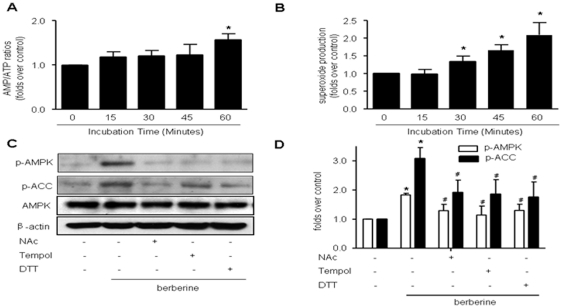
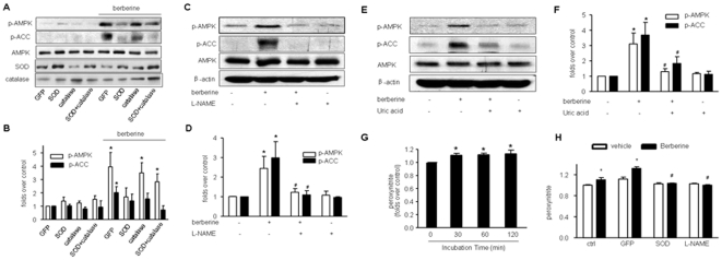
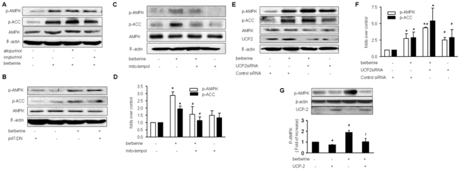
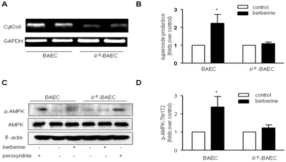
In BAEC, Berberine caused a dose- and time-dependent increase in the phosphorylation of AMPK at Thr172 and acetyl CoA carboxylase (ACC) at Ser79, a well characterized downstream target of AMPK. Concomitantly, Berberine increased peroxynitrite, a potent oxidant formed by simultaneous generation of superoxide and nitric oxide. Pre-incubation of BAEC with anti-oxidants markedly attenuated Berberine-enhanced phosphorylation of both AMPK and ACC. Consistently, adenoviral expression of superoxide dismutase and pretreatment of L-N(G)-Nitroarginine methyl ester (L-NAME; a non-selective NOS inhibitor) blunted Berberine-induced phosphorylation of AMPK. Furthermore, mitochondria-targeted tempol (mito-tempol) pretreatment or expression of uncoupling protein attenuated AMPK activation caused by Berberine. Depletion of mitochondria abolished the effects of Berberine on AMPK in EC. Finally, Berberine significantly increased the phosphorylation of LKB1 at Ser307 and gene silencing of LKB1 attenuated Berberine-enhanced AMPK Thr172 phosphorylation in BAEC.
Our results suggest that mitochondria-derived superoxide anions and peroxynitrite are required for Berberine-induced AMPK activation in endothelial cells.
氧化还原状态是细胞功能的一个关键决定因素,任何重大失衡都可能导致严重的损伤或死亡。
本研究旨在确定 AMP 激活的蛋白激酶(AMPK),一种细胞能量传感器,是否被小檗碱在血管内皮细胞(EC)中产生的氧化剂激活。
将牛主动脉内皮细胞(BAEC)暴露于小檗碱中。孵育后监测 AMPK 活性和活性氧的产生。
在 BAEC 中,小檗碱引起 AMPK 在 Thr172 处和乙酰辅酶 A 羧化酶(ACC)在 Ser79 处的磷酸化呈剂量和时间依赖性增加,这是 AMPK 的一个很好的下游靶点。同时,小檗碱增加了过氧亚硝酸盐,这是一种由超氧化物和一氧化氮同时产生的强氧化剂。BAEC 用抗氧化剂预孵育可显著减弱小檗碱增强的 AMPK 和 ACC 的磷酸化。一致地,过氧化物歧化酶的腺病毒表达和 L-N(G)-硝基精氨酸甲酯(L-NAME;一种非选择性 NOS 抑制剂)预处理减弱了小檗碱诱导的 AMPK 磷酸化。此外,线粒体靶向的tempo(mito-tempol)预处理或解偶联蛋白的表达减弱了小檗碱引起的 AMPK 激活。线粒体耗竭消除了小檗碱对 EC 中 AMPK 的影响。最后,小檗碱显著增加了 LKB1 在 Ser307 的磷酸化,并且 LKB1 的基因沉默减弱了 BAEC 中小檗碱增强的 AMPK Thr172 磷酸化。
我们的结果表明,线粒体衍生的超氧阴离子和过氧亚硝酸盐是小檗碱诱导内皮细胞 AMPK 激活所必需的。