Department of Cell Research and Immunology, The George S Wise Faculty of Life Sciences, Tel Aviv University, Tel Aviv, Israel.
PLoS Pathog. 2010 Nov 11;6(11):e1001183. doi: 10.1371/journal.ppat.1001183.
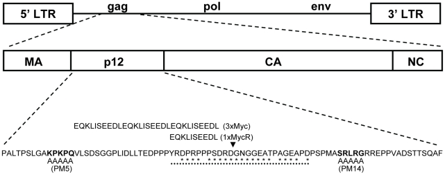
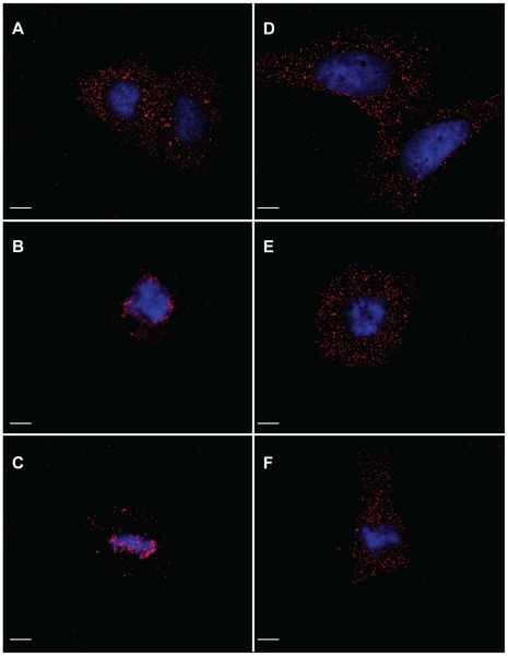
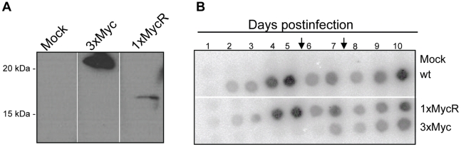
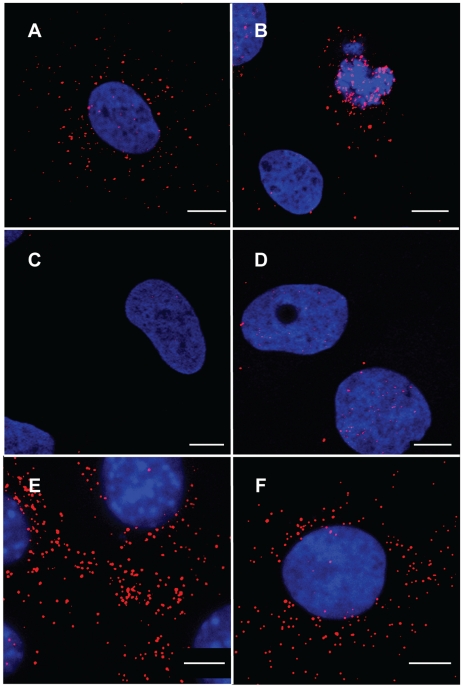
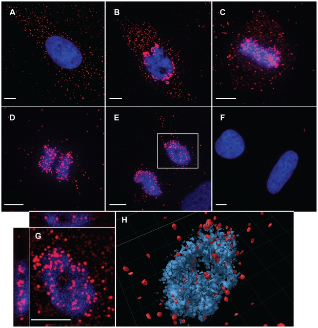
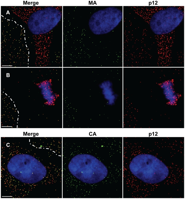
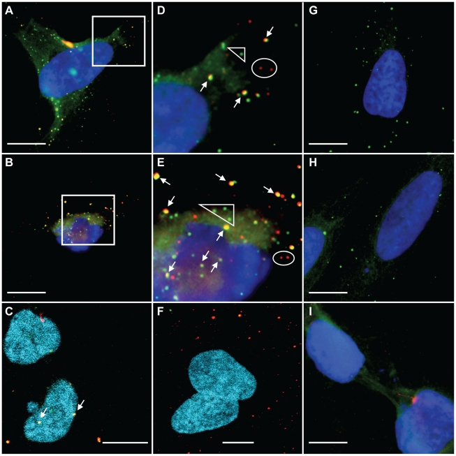
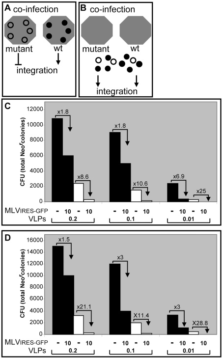
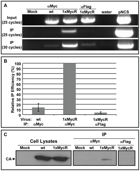
The p12 protein is a cleavage product of the Gag precursor of the murine leukemia virus (MLV). Specific mutations in p12 have been described that affect early stages of infection, rendering the virus replication-defective. Such mutants showed normal generation of genomic DNA but no formation of circular forms, which are markers of nuclear entry by the viral DNA. This suggested that p12 may function in early stages of infection but the precise mechanism of p12 action is not known. To address the function and follow the intracellular localization of the wt p12 protein, we generated tagged p12 proteins in the context of a replication-competent virus, which allowed for the detection of p12 at early stages of infection by immunofluorescence. p12 was found to be distributed to discrete puncta, indicative of macromolecular complexes. These complexes were localized to the cytoplasm early after infection, and thereafter accumulated adjacent to mitotic chromosomes. This chromosomal accumulation was impaired for p12 proteins with a mutation that rendered the virus integration-defective. Immunofluorescence demonstrated that intracellular p12 complexes co-localized with capsid, a known constituent of the MLV pre-integration complex (PIC), and immunofluorescence combined with fluorescent in situ hybridization (FISH) revealed co-localization of the p12 proteins with the incoming reverse transcribed viral DNA. Interactions of p12 with the capsid and with the viral DNA were also demonstrated by co-immunoprecipitation. These results imply that p12 proteins are components of the MLV PIC. Furthermore, a large excess of wt PICs did not rescue the defect in integration of PICs derived from mutant p12 particles, demonstrating that p12 exerts its function as part of this complex. Altogether, these results imply that p12 proteins are constituent of the MLV PIC and function in directing the PIC from the cytoplasm towards integration.
p12 蛋白是鼠白血病病毒(MLV)Gag 前体的裂解产物。已经描述了影响感染早期阶段的 p12 中的特定突变,使病毒复制缺陷。这些突变体显示出基因组 DNA 的正常生成,但没有形成环状形式,这是病毒 DNA 进入核的标志物。这表明 p12 可能在感染的早期阶段发挥作用,但 p12 作用的确切机制尚不清楚。为了研究功能并跟踪 wt p12 蛋白的细胞内定位,我们在复制有效的病毒背景下生成了标记的 p12 蛋白,这允许通过免疫荧光在感染的早期阶段检测到 p12。发现 p12 分布在离散的小点上,表明存在大分子复合物。这些复合物在感染后早期定位于细胞质中,然后在有丝分裂染色体附近积累。对于使病毒整合缺陷的 p12 蛋白,这种染色体积累受到损害。免疫荧光显示细胞内 p12 复合物与衣壳共定位,衣壳是 MLV 预整合复合物(PIC)的已知成分,免疫荧光结合荧光原位杂交(FISH)显示 p12 蛋白与传入的逆转录病毒 DNA 共定位。p12 与衣壳和病毒 DNA 的相互作用也通过共免疫沉淀得到证实。这些结果表明 p12 蛋白是 MLV PIC 的组成部分。此外,大量 wt PIC 不能挽救源自突变 p12 颗粒的 PIC 整合缺陷,表明 p12 作为该复合物的一部分发挥其功能。总之,这些结果表明 p12 蛋白是 MLV PIC 的组成部分,并在将 PIC 从细胞质引导至整合中发挥作用。