Schwartzkopff J, Bredow L, Mahlenbrey S, Boehringer D, Reinhard T
University Eye Hospital Freiburg, Killianstr. 5, 79106 Freiburg, Germany.
Mol Vis. 2010 Nov 11;16:2368-75.
Corneal endothelial cells (EC) are crucial for maintaining corneal clarity before and after keratoplasty. Since it is thought that corneal graft rejection leads to irreversible EC loss and transplant failure, we quantified immune mediated EC loss in the rat keratoplasty model and analyzed whether the EC layer would then regenerate.
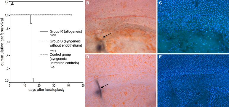
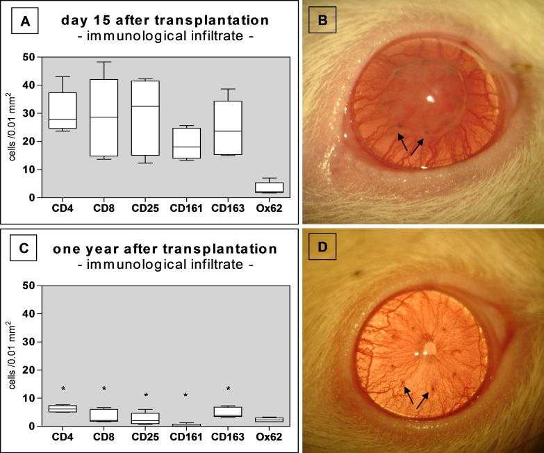
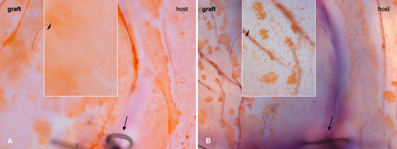
Rats were subjected to orthotopic penetrating keratoplasty. We compared endothelial responses to immunological EC loss following allogeneic transplantations between Fisher and Lewis rats (group R) to those following mechanical EC removal in a syngeneic setting between Lewis rats (group S). Animals were followed clinically for corneal opacity for up to one year. Bulbi were excised and prepared for histological examination at different time points: ECs were defined and characterized using Alicarin red S/ DAPI staining on corneal flatmounts. Ki-67/ DAPI staining on flatmount preparations served to detect cell proliferation. Immunohistochemical staining of corneal cryosections was used to characterize infiltrating immune cells.
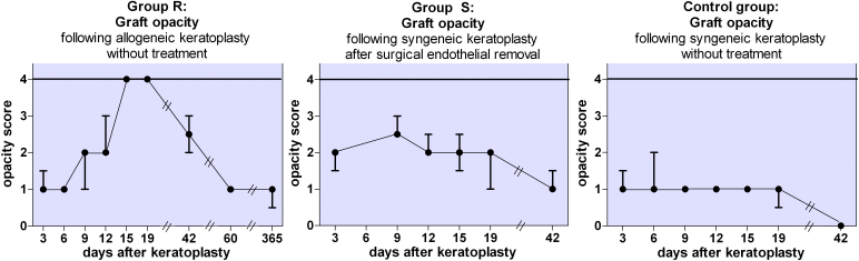
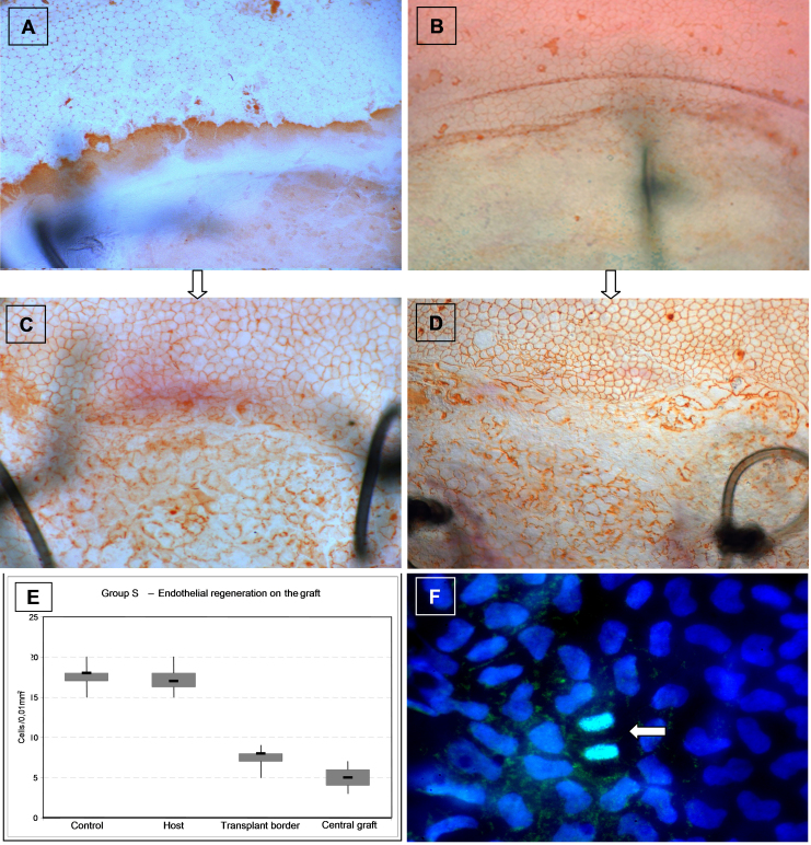
GROUP R: After about two weeks the allografts were completely opaque, which was accompanied by a massive leukocyte infiltration in conjunction with EC destruction, signifying rejection. EC loss without an immune reaction (group S) resulted only in medium opacity levels. In both groups, all grafts regained clarity in the following weeks to months, and a newly-formed endothelial cell layer with irregular and enlarged ECs became apparent on the formerly EC free grafts. Scattered Ki-67 positive cells within the endothelial cell layer were observed during re-endothelialization. In addition to re-endothelialization, the immunological infiltration seen in the allografts at the time of rejection had subsided after one year.
Re-endothelialization following keratoplasty takes place in the rat in vivo and restores graft clarity, following both immunological or surgical destruction of ECs. Following rejection, EC replacement is accompanied by a reduction of immune infiltrates. Peripheral recipient ECs are a sufficient source for graft re-endothelialization, as seen in rats following EC removal. Our results suggest that ECs both proliferate and enlarge during re-endothelialization in the rat keratoplasty model.
角膜内皮细胞(EC)对于维持角膜移植术前和术后的角膜透明度至关重要。由于人们认为角膜移植排斥会导致不可逆的EC损失和移植失败,我们在大鼠角膜移植模型中对免疫介导的EC损失进行了量化,并分析了EC层随后是否会再生。
对大鼠进行原位穿透性角膜移植。我们比较了Fisher大鼠和Lewis大鼠之间同种异体移植后免疫性EC损失的内皮反应(R组)与Lewis大鼠之间同基因环境下机械性EC去除后的内皮反应(S组)。对动物进行临床随访,观察角膜混浊情况长达一年。在不同时间点切除眼球并准备进行组织学检查:使用阿利卡林红S/ DAPI染色在角膜平铺标本上对EC进行定义和表征。在平铺标本上进行Ki-67/ DAPI染色用于检测细胞增殖。角膜冰冻切片的免疫组织化学染色用于表征浸润的免疫细胞。
R组:大约两周后,同种异体移植物完全混浊,伴有大量白细胞浸润和EC破坏,表明发生排斥反应。无免疫反应的EC损失(S组)仅导致中度混浊水平。在两组中,所有移植物在接下来的数周数月内恢复了透明度,并且在先前无EC的移植物上出现了由不规则且增大的EC组成的新形成的内皮细胞层。在再内皮化过程中,在内皮细胞层内观察到散在的Ki-67阳性细胞。除了再内皮化外,排斥时同种异体移植物中所见的免疫浸润在一年后已消退。
角膜移植术后的再内皮化在大鼠体内发生,并在EC受到免疫或手术破坏后恢复移植物透明度。排斥反应后,EC替代伴随着免疫浸润的减少。外周受体EC是移植物再内皮化的充足来源,如在大鼠EC去除后所见。我们的结果表明,在大鼠角膜移植模型中,EC在再内皮化过程中既增殖又增大。