Madias John E
Mount Sinai School of Medicine of the New York University, and the Division of Cardiology, Elmhurst Hospital Center, New York, NY.
Indian Pacing Electrophysiol J. 2010 Oct 31;10(10):464-73.
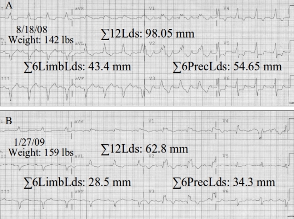
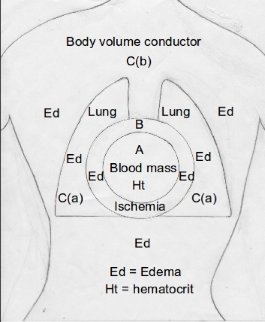
A 3-compartment mechanistic model is proposed to explain the attenuation of the electrocardiographic QRS complexes (↓QRSV) observed in patients with heart failure (HF). This includes the effects of increased intracardiac blood volume and decreased hematocrit due to blood dilution (1st compartment), the heart's alteration in electrogenesis due to possible ischemia or inflammation, leading to myocardial edema, (2nd compartment), and the passive volume conductor of the tissue and organ constituents of the thorax and the entire body, with their resistivity changes due to increased fluid content (pulmonary and peripheral edema) (3rd compartment). The clinical implications of the model are outlined.
提出了一种三室机制模型来解释心力衰竭(HF)患者中观察到的心电图QRS波群衰减(↓QRSV)。这包括由于血液稀释导致的心内血容量增加和血细胞比容降低的影响(第一室)、由于可能的缺血或炎症导致心脏电发生改变从而引起心肌水肿的影响(第二室),以及胸部和整个身体的组织和器官成分的被动容积导体,其电阻率因液体含量增加(肺和外周水肿)而发生变化(第三室)。概述了该模型的临床意义。