Department of Biochemistry and Molecular Genetics, University of Illinois at Chicago College of Medicine, 900 S. Ashland Avenue, Chicago, IL 60607, USA.
Adv Exp Med Biol. 2011;691:595-603. doi: 10.1007/978-1-4419-6612-4_63.
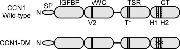
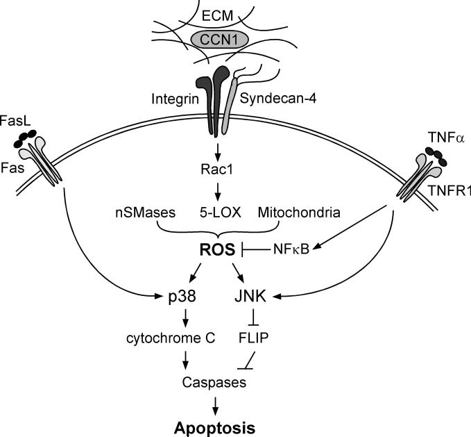
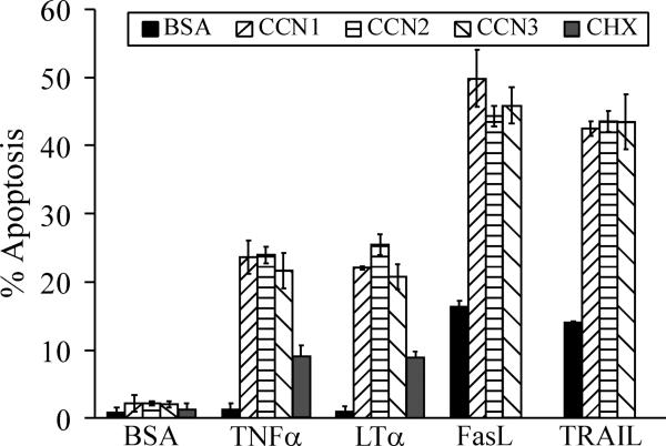
It has long been appreciated that the apoptotic activity of TNFα is context-dependent, and requires inhibition of NFκB signaling or protein synthesis to be manifested in most normal cells in culture. Recent studies have uncovered an unexpected pro-apoptotic synergism between TNF cytokines and the CCN family of extracellular matrix proteins, which are dynamically expressed at sites of injury repair and inflammation. The presence of CCN1, CCN2, or CCN3 allows TNFα to induce apoptosis with high efficacy without perturbation of NFκB signaling or protein synthesis, thus converting TNFα from a proliferation-promoting protein into an apoptotic inducer. CCN proteins also enhance the cytotoxicity of other TNF family cytokines including LTα, FasL, and TRAIL. CCN proteins synergize with TNF cytokines through binding to integrin αβ and the heparan sulfate proteoglycan (HSPG) syndecan-4 to induce reactive oxygen species (ROS) accumulation. Knockin mice that express a CCN1 mutant defective for binding αβ-HSPG are severely blunted in TNFα- and Fas-mediated apoptosis, indicating that CCN1 is a physiologic regulator of these processes. Thus, CCN proteins in the extracellular matrix microenvironment can provide the contextual cues for the cytotoxicity of TNFα and related cytokines, and profoundly influence their activity.
长期以来,人们一直认为 TNFα 的凋亡活性是依赖于上下文的,并且需要抑制 NFκB 信号或蛋白质合成才能在大多数正常培养细胞中表现出来。最近的研究揭示了 TNF 细胞因子与细胞外基质蛋白 CCN 家族之间一种出乎意料的促凋亡协同作用,这些蛋白在损伤修复和炎症部位动态表达。CCN1、CCN2 或 CCN3 的存在允许 TNFα 在不干扰 NFκB 信号或蛋白质合成的情况下高效诱导细胞凋亡,从而将 TNFα 从促进增殖的蛋白转化为凋亡诱导剂。CCN 蛋白还增强了其他 TNF 家族细胞因子(包括 LTα、FasL 和 TRAIL)的细胞毒性。CCN 蛋白通过与整合素 αβ 和硫酸乙酰肝素蛋白聚糖 (HSPG) 连接蛋白聚糖-4 结合,与 TNF 细胞因子协同作用,诱导活性氧 (ROS) 积累。表达一种 CCN1 突变体的嵌合小鼠,该突变体不能与 αβ-HSPG 结合,在 TNFα 和 Fas 介导的凋亡中严重受损,表明 CCN1 是这些过程的生理调节剂。因此,细胞外基质微环境中的 CCN 蛋白可以为 TNFα 和相关细胞因子的细胞毒性提供上下文线索,并深刻影响它们的活性。