Department of Health Sciences, University of Jyväskylä, Finland.
Nutr Metab (Lond). 2011 Jan 17;8(1):1. doi: 10.1186/1743-7075-8-1.
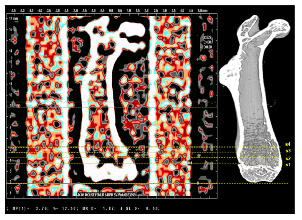
Obesity and osteoporosis, two possibly related conditions, are rapidly expanding health concerns in modern society. Both of them are associated with sedentary life style and nutrition. To investigate the effects of diet-induced obesity and voluntary physical activity we used high resolution micro-computed tomography (μCT) together with peripheral quantitative computed tomography (pQCT) to examine the microstructure of the distal femoral metaphysis in mice.
Forty 7-week-old male C57BL/6J mice were assigned to 4 groups: control (C), control + running (CR), high-fat diet (HF), and high-fat diet + running (HFR). After a 21-week intervention, all the mice were sacrificed and the left femur dissected for pQCT and μCT measurements.
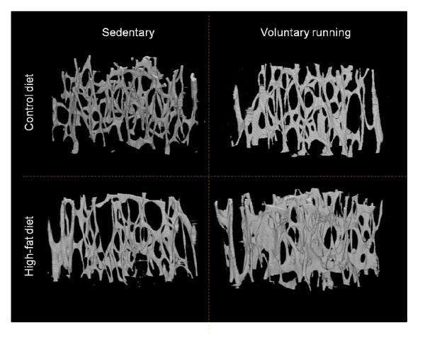
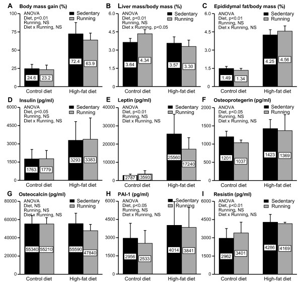
The mice fed the high-fat diet showed a significant weight gain (over 70% for HF and 60% for HFR), with increased epididymal fat pad mass and impaired insulin sensitivity. These obese mice had significantly higher trabecular connectivity density, volume, number, thickness, area and mass, and smaller trabecular separation. At the whole bone level, they had larger bone circumference and cross-sectional area and higher density-weighted maximal, minimal, and polar moments of inertia. Voluntary wheel running decreased all the cortical bone parameters, but increased the trabecular mineral density, and decreased the pattern factor and structure model index towards a more plate-like structure.
The results suggest that in mice the femur adapts to obesity by improving bone strength both at the whole bone and micro-structural level. Adaptation to running exercise manifests itself in increased trabecular density and improved 3D structure, but in a limited overall bone growth.
肥胖症和骨质疏松症是现代社会中迅速扩大的健康问题,二者可能与久坐的生活方式和营养有关。为了研究饮食诱导肥胖和自愿体力活动的影响,我们使用高分辨率微计算机断层扫描(μCT)和外周定量计算机断层扫描(pQCT)检查了小鼠远端股骨干骺端的微观结构。
将 40 只 7 周龄雄性 C57BL/6J 小鼠分为 4 组:对照组(C)、对照组+跑步(CR)、高脂肪饮食组(HF)和高脂肪饮食+跑步(HFR)。经过 21 周的干预后,所有小鼠被处死,取左侧股骨进行 pQCT 和 μCT 测量。
高脂饮食组的小鼠体重明显增加(HF 组增加超过 70%,HFR 组增加超过 60%),附睾脂肪垫质量增加,胰岛素敏感性受损。这些肥胖小鼠的小梁连接密度、体积、数量、厚度、面积和质量显著增加,而小梁分离度显著减小。在整个骨水平,它们的骨周长和横截面积更大,密度加权的最大、最小和极惯性矩更高。自愿轮跑降低了所有皮质骨参数,但增加了小梁骨密度,降低了模式因子和结构模型指数,向更板状结构发展。
这些结果表明,在肥胖小鼠中,股骨通过改善整体骨和微观结构水平的骨强度来适应肥胖。对跑步运动的适应表现为小梁密度增加和 3D 结构改善,但总体骨生长有限。