Calkins Marcus J, Reddy P Hemachandra
Neurogenetics Laboratory, Division of Neuroscience, Oregon National Primate Research Center, Oregon Health & Science University, 505 NW 185th Avenue, Beaverton, OR 97006, USA.
Biochim Biophys Acta. 2011 Apr;1812(4):507-13. doi: 10.1016/j.bbadis.2011.01.007. Epub 2011 Jan 15.
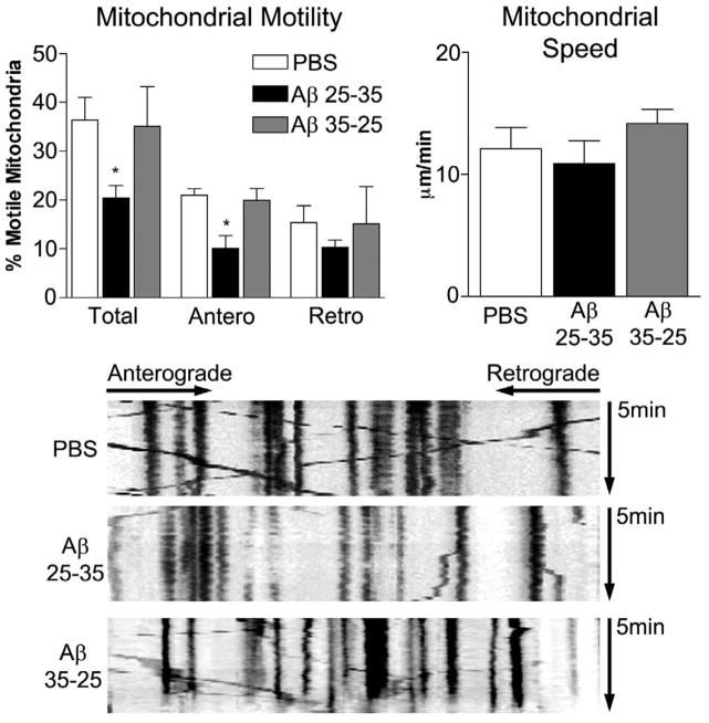
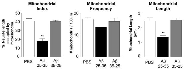
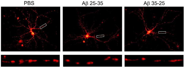
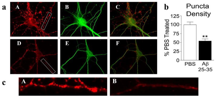
Loss of synapses and synaptic damage are the best correlates of cognitive decline identified in patients with Alzheimer's disease (AD), and mitochondrial oxidative damage and synaptic pathology have been identified as early events in the progression of AD. The progressive accumulation of amyloid beta (Aβ) in synapses and synaptic mitochondria are hypothesized to cause synaptic degeneration and cognitive decline in patients with AD. However, the precise mechanistic link between Aβ and mitochondria is not well understood. The purpose of this study was to better understand the effects of Aβ on mitochondrial axonal transport and synaptic alterations in AD. Using mouse hippocampal neurons and Aβ(25-35) peptide, we studied axonal transport of mitochondria, including mitochondrial motility, mitochondrial length and size, mitochondrial index per neurite, and synaptic alterations of the hippocampal neurons. In the PBS-treated neurons, 36.4±4.7% of the observed mitochondria were motile, with 21.0±1.3% moving anterograde and 15.4±3.4% moving retrograde and the average speed of movement was 12.1±1.8μm/min. In contrast, in the Aβ-treated neurons, the number of motile mitochondria were significantly less, at 20.4±2.6% (P<0.032), as were those moving anterograde (10.1±2.6%, P<0.016) relative to PBS-treated neurons, suggesting that the Aβ(25-35) peptide impairs axonal transport of mitochondria in AD neurons. In the Aβ-treated neurons, the average speed of motile mitochondria was also less, at 10.9±1.9μm/min, and mitochondrial length was significantly decreased. Further, synaptic immunoreactivity was also significantly less in the Aβ-treated neurons relative to the PBS-treated neurons, indicating that Aβ affects synaptic viability. These findings suggest that, in neurons affected by AD, Aβ is toxic, impairs mitochondrial movements, reduces mitochondrial length, and causes synaptic degeneration.
突触丧失和突触损伤是阿尔茨海默病(AD)患者认知功能下降的最佳相关因素,线粒体氧化损伤和突触病理已被确定为AD进展过程中的早期事件。淀粉样β蛋白(Aβ)在突触和突触线粒体中的逐渐积累被认为是导致AD患者突触退化和认知功能下降的原因。然而,Aβ与线粒体之间的确切机制联系尚不清楚。本研究的目的是更好地了解Aβ对AD中线粒体轴突运输和突触改变的影响。使用小鼠海马神经元和Aβ(25-35)肽,我们研究了线粒体的轴突运输,包括线粒体运动性、线粒体长度和大小、每个神经突的线粒体指数以及海马神经元的突触改变。在PBS处理的神经元中,观察到的线粒体中有36.4±4.7%是可移动的,其中21.0±1.3%向前移动,15.4±3.4%向后移动,平均移动速度为12.1±1.8μm/分钟。相比之下,在Aβ处理的神经元中,可移动线粒体的数量显著减少,为20.4±2.6%(P<0.032),向前移动的线粒体数量(10.1±2.6%,P<0.016)也相对于PBS处理的神经元减少,这表明Aβ(25-35)肽损害了AD神经元中线粒体的轴突运输。在Aβ处理的神经元中,可移动线粒体的平均速度也较低,为10.9±1.9μm/分钟,线粒体长度显著缩短。此外,与PBS处理的神经元相比,Aβ处理的神经元中的突触免疫反应性也显著降低,表明Aβ影响突触活力。这些发现表明,在受AD影响的神经元中,Aβ具有毒性,损害线粒体运动,缩短线粒体长度,并导致突触退化。