• 文献检索
  • 文档翻译
  • 深度研究
  • 学术资讯
  • Suppr Zotero 插件Zotero 插件
  • 邀请有礼
  • 套餐&价格
  • 历史记录
应用&插件
Suppr Zotero 插件Zotero 插件浏览器插件Mac 客户端Windows 客户端微信小程序
定价
高级版会员购买积分包购买API积分包
服务
文献检索文档翻译深度研究API 文档MCP 服务
关于我们
关于 Suppr公司介绍联系我们用户协议隐私条款
关注我们

Suppr 超能文献

核心技术专利:CN118964589B侵权必究
粤ICP备2023148730 号-1Suppr @ 2026

文献检索

告别复杂PubMed语法,用中文像聊天一样搜索,搜遍4000万医学文献。AI智能推荐,让科研检索更轻松。

立即免费搜索

文件翻译

保留排版,准确专业,支持PDF/Word/PPT等文件格式,支持 12+语言互译。

免费翻译文档

深度研究

AI帮你快速写综述,25分钟生成高质量综述,智能提取关键信息,辅助科研写作。

立即免费体验

肝硬化中的肝细胞结节:血流动力学评估(血管造影辅助CT),特别提及多步骤肝癌发生过程

Hepatocelluar nodules in liver cirrhosis: hemodynamic evaluation (angiography-assisted CT) with special reference to multi-step hepatocarcinogenesis.

作者信息

Matsui Osamu, Kobayashi Satoshi, Sanada Junichiro, Kouda Wataru, Ryu Yasuji, Kozaka Kazuto, Kitao Azusa, Nakamura Koichi, Gabata Toshifumi

机构信息

Department of Radiology, Graduate School of Medical Science, Kanazawa University, Japan.

出版信息

Abdom Imaging. 2011 Jun;36(3):264-72. doi: 10.1007/s00261-011-9685-1.

DOI:10.1007/s00261-011-9685-1
PMID:21267562
原文链接:https://pmc.ncbi.nlm.nih.gov/articles/PMC3102849/
Abstract

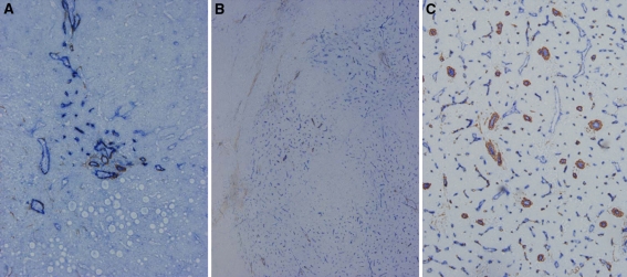
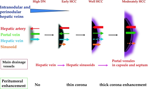
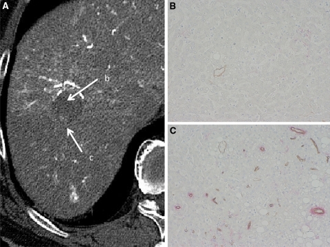
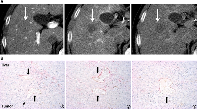
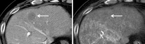
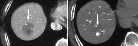
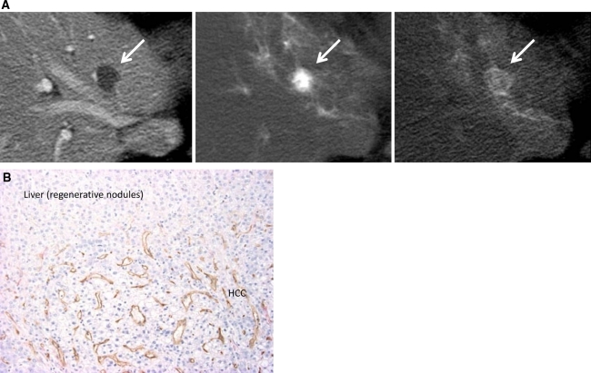
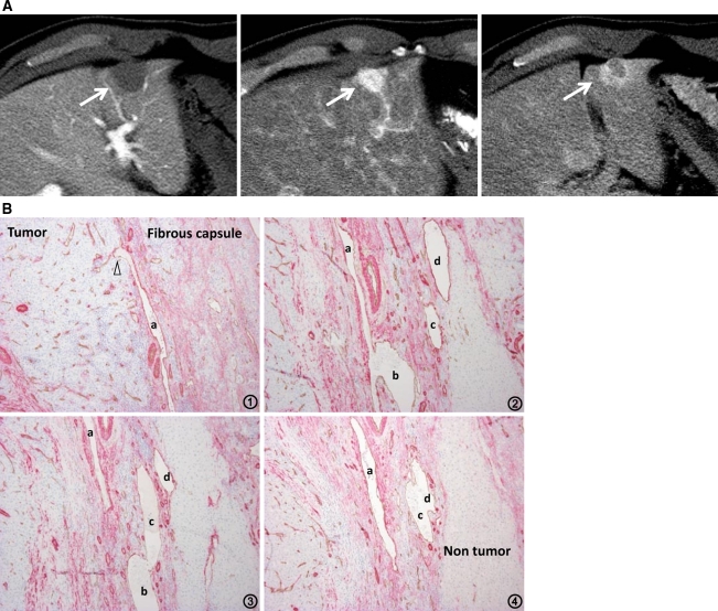
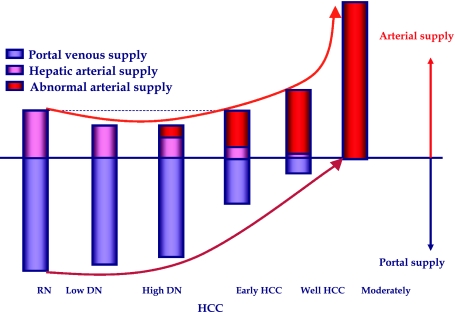
To understand the hemodynamics of hepatocellular carcinoma (HCC) is important for the precise imaging diagnosis and treatment, because there is an intense correlation between their hemodynamics and pathophysiology. Angiogenesis such as sinusoidal capillarization and unpaired arteries shows gradual increase during multi-step hepatocarcinogenesis from high-grade dysplastic nodule to classic hypervascular HCC. In accordance with this angiogenesis, the intranodular portal supply is decreased, whereas the intranodular arterial supply is first decreased during the early stage of hepatocarcinogenesis and then increased in parallel with increasing grade of malignancy of the nodules. On the other hand, the main drainage vessels of hepatocellular nodules change from hepatic veins to hepatic sinusoids and then to portal veins during multi-step hepatocarcinogenesis, mainly due to disappearance of the hepatic veins from the nodules. Therefore, in early HCC, no perinodular corona enhancement is seen on portal to equilibrium phase CT, but it is definite in hypervascular classical HCC. Corona enhancement is thicker in encapsulated HCC and thin in HCC without pseudocapsule. To understand these hemodynamic changes during multi-step hepatocarcinogenesis is important, especially for early diagnosis and treatment of HCCs.

摘要

了解肝细胞癌(HCC)的血流动力学对于精确的影像诊断和治疗至关重要,因为其血流动力学与病理生理学之间存在密切关联。在从高级别发育异常结节到经典高血供HCC的多步骤肝癌发生过程中,诸如窦状毛细血管化和不成对动脉等血管生成呈逐渐增加趋势。与这种血管生成相一致,结节内门静脉供血减少,而结节内动脉供血在肝癌发生早期首先减少,然后随着结节恶性程度的增加而平行增加。另一方面,在多步骤肝癌发生过程中,肝细胞结节的主要引流血管从肝静脉变为肝血窦,然后变为门静脉,这主要是由于结节内肝静脉消失所致。因此,在早期HCC中,门静脉期至平衡期CT上未见结节周围晕圈强化,但在高血供经典HCC中则明确可见。包膜型HCC的晕圈强化较厚,无假包膜的HCC的晕圈强化较薄。了解多步骤肝癌发生过程中的这些血流动力学变化非常重要,尤其是对于HCC的早期诊断和治疗。

https://cdn.ncbi.nlm.nih.gov/pmc/blobs/d156/3102849/6efa46b40ae5/261_2011_9685_Fig9_HTML.jpg
https://cdn.ncbi.nlm.nih.gov/pmc/blobs/d156/3102849/d506f366c5f2/261_2011_9685_Fig1_HTML.jpg
https://cdn.ncbi.nlm.nih.gov/pmc/blobs/d156/3102849/ddbbd59f17c3/261_2011_9685_Fig2_HTML.jpg
https://cdn.ncbi.nlm.nih.gov/pmc/blobs/d156/3102849/4767540af8c3/261_2011_9685_Fig3_HTML.jpg
https://cdn.ncbi.nlm.nih.gov/pmc/blobs/d156/3102849/2ba203d22c47/261_2011_9685_Fig4_HTML.jpg
https://cdn.ncbi.nlm.nih.gov/pmc/blobs/d156/3102849/d99f7473a433/261_2011_9685_Fig5_HTML.jpg
https://cdn.ncbi.nlm.nih.gov/pmc/blobs/d156/3102849/6e992d05b0e9/261_2011_9685_Fig6_HTML.jpg
https://cdn.ncbi.nlm.nih.gov/pmc/blobs/d156/3102849/1f69d37db94a/261_2011_9685_Fig7_HTML.jpg
https://cdn.ncbi.nlm.nih.gov/pmc/blobs/d156/3102849/5109d87e20f2/261_2011_9685_Fig8_HTML.jpg
https://cdn.ncbi.nlm.nih.gov/pmc/blobs/d156/3102849/6efa46b40ae5/261_2011_9685_Fig9_HTML.jpg

相似文献

1
Hepatocelluar nodules in liver cirrhosis: hemodynamic evaluation (angiography-assisted CT) with special reference to multi-step hepatocarcinogenesis.肝硬化中的肝细胞结节:血流动力学评估(血管造影辅助CT),特别提及多步骤肝癌发生过程
Abdom Imaging. 2011 Jun;36(3):264-72. doi: 10.1007/s00261-011-9685-1.
2
Hepatocarcinogenesis: multistep changes of drainage vessels at CT during arterial portography and hepatic arteriography--radiologic-pathologic correlation.肝癌发生:动脉门静脉造影和肝动脉造影期间CT上引流血管的多步变化——放射学与病理学相关性
Radiology. 2009 Aug;252(2):605-14. doi: 10.1148/radiol.2522081414.
3
Imaging of multistep human hepatocarcinogenesis by CT during intra-arterial contrast injection.肝动脉内注射造影剂时CT对人肝癌多步骤发生过程的成像
Intervirology. 2004;47(3-5):271-6. doi: 10.1159/000078478.
4
Atypical large well-differentiated hepatocellular carcinoma with benign nature: a new clinical entity.具有良性特征的非典型高分化肝细胞癌:一种新的临床实体。
Intervirology. 2004;47(3-5):227-37. doi: 10.1159/000078475.
5
Correlation between the blood supply and grade of malignancy of hepatocellular nodules associated with liver cirrhosis: evaluation by CT during intraarterial injection of contrast medium.肝硬化相关肝细胞结节的血供与恶性程度之间的相关性:动脉内注射造影剂时CT评估
AJR Am J Roentgenol. 1999 Apr;172(4):969-76. doi: 10.2214/ajr.172.4.10587130.
6
Sequential hemodynamic change in hepatocellular carcinoma and dysplastic nodules: CT angiography and pathologic correlation.肝细胞癌和发育异常结节的序贯血流动力学变化:CT血管造影与病理对照
AJR Am J Roentgenol. 2002 Apr;178(4):885-97. doi: 10.2214/ajr.178.4.1780885.
7
Progression to hypervascular hepatocellular carcinoma: correlation with intranodular blood supply evaluated with CT during intraarterial injection of contrast material.进展为富血管性肝细胞癌:与动脉内注射造影剂期间CT评估的结节内血供的相关性
Radiology. 2002 Oct;225(1):143-9. doi: 10.1148/radiol.2251011298.
8
Imaging of early hepatocellular carcinoma and adenomatous hyperplasia (dysplastic nodules) with dynamic ct and a combination of CT and angiography: experience with resected liver specimens.早期肝细胞癌和腺瘤样增生(发育异常结节)的动态CT成像以及CT与血管造影相结合的成像:对切除肝脏标本的研究经验
Intervirology. 2004;47(3-5):199-208. doi: 10.1159/000078473.
9
Vascular changes in hepatocellular carcinoma: correlation of radiologic and pathologic findings.肝细胞癌中的血管变化:放射学与病理学发现的相关性
AJR Am J Roentgenol. 1999 Nov;173(5):1213-7. doi: 10.2214/ajr.173.5.10541091.
10
Hemodynamics and progression of a hypervascular focus in a borderline lesion of hepatocellular carcinoma: analysis by angiography-assisted CT and histopathology.血管造影辅助 CT 与组织病理学分析肝细胞癌边界性病变中富血管病灶的血液动力学变化与进展。
Jpn J Radiol. 2014 Feb;32(2):69-79. doi: 10.1007/s11604-013-0268-7. Epub 2013 Dec 12.

引用本文的文献

1
Optimizing Imaging Parameters for Assessment of Hepatocellular Carcinoma Using Photon-Counting Detector Computed Tomography-Impact of Reconstruction Kernel and Slice Thickness.使用光子计数探测器计算机断层扫描优化评估肝细胞癌的成像参数——重建核和层厚的影响
Tomography. 2025 Jun 27;11(7):77. doi: 10.3390/tomography11070077.
2
The evolution of anatomical hepatectomy: Past, present, and future.解剖性肝切除术的演变:过去、现在与未来。
ILIVER. 2022 Oct 11;1(3):199-204. doi: 10.1016/j.iliver.2022.09.003. eCollection 2022 Sep.
3
Immunoscore Predicted by Dynamic Contrast-Enhanced Computed Tomography Can Be a Non-Invasive Biomarker for Immunotherapy Susceptibility of Hepatocellular Carcinoma.

本文引用的文献

1
Hepatocarcinogenesis: multistep changes of drainage vessels at CT during arterial portography and hepatic arteriography--radiologic-pathologic correlation.肝癌发生:动脉门静脉造影和肝动脉造影期间CT上引流血管的多步变化——放射学与病理学相关性
Radiology. 2009 Aug;252(2):605-14. doi: 10.1148/radiol.2522081414.
2
Pathologic diagnosis of early hepatocellular carcinoma: a report of the international consensus group for hepatocellular neoplasia.早期肝细胞癌的病理诊断:肝细胞肿瘤国际共识小组报告
Hepatology. 2009 Feb;49(2):658-64. doi: 10.1002/hep.22709.
3
Imaging of benign hypervascular hepatocellular nodules in alcoholic liver cirrhosis: differentiation from hypervascular hepatocellular carcinoma.
动态对比增强计算机断层扫描预测的免疫评分可作为肝细胞癌免疫治疗敏感性的非侵入性生物标志物。
Cancers (Basel). 2025 Mar 11;17(6):948. doi: 10.3390/cancers17060948.
4
Comparing health insurance-reimbursed lenvatinib and self-paid atezolizumab plus bevacizumab in patients with unresectable hepatocellular carcinoma.比较医保报销的乐伐替尼与自费的阿替利珠单抗联合贝伐单抗用于不可切除肝细胞癌患者的情况。
Am J Cancer Res. 2025 Feb 15;15(2):811-823. doi: 10.62347/BYIE2654. eCollection 2025.
5
Molecular Classification of Hepatocellular Carcinoma based on Zoned Metabolic Feature and Oncogenic Signaling Pathway.基于分区代谢特征和致癌信号通路的肝细胞癌分子分类
Clin Mol Hepatol. 2025 Mar 11. doi: 10.3350/cmh.2024.1088.
6
Early Enhancement in Contrast-Enhanced Computed Tomography Is an Index of , , , and Expression in Canine Hepatocellular Carcinoma: A Preliminary Study.对比增强计算机断层扫描中的早期强化是犬肝细胞癌中、、、和表达的一个指标:一项初步研究。
Vet Sci. 2025 Feb 7;12(2):137. doi: 10.3390/vetsci12020137.
7
Correlation Between Dynamic Contrast-Enhanced CT Imaging Signs and Differentiation Grade and Microvascular Invasion of Hepatocellular Carcinoma.动态对比增强CT成像征象与肝细胞癌分化程度及微血管侵犯的相关性
J Hepatocell Carcinoma. 2025 Jan 8;12:1-14. doi: 10.2147/JHC.S489387. eCollection 2025.
8
Radiological Assessment and Therapeutic Evaluation in Hepatocellular Carcinoma: Differentiation and Treatment Response with Japanese Guidelines.肝细胞癌的放射学评估与治疗评价:依据日本指南进行的鉴别与治疗反应分析
Cancers (Basel). 2024 Dec 31;17(1):101. doi: 10.3390/cancers17010101.
9
Hepatocellular carcinoma: updates on epidemiology, surveillance, diagnosis and treatment.肝细胞癌:流行病学、监测、诊断与治疗的最新进展
Clin Mol Hepatol. 2025 Feb;31(Suppl):S228-S254. doi: 10.3350/cmh.2024.0824. Epub 2024 Dec 26.
10
Evaluating the role of quantitative computed tomography perfusion parameters in differentiating hepatocellular carcinoma from other hepatic neoplasms.评估定量计算机断层扫描灌注参数在鉴别肝细胞癌与其他肝脏肿瘤中的作用。
Abdom Radiol (NY). 2025 Jun;50(6):2440-2452. doi: 10.1007/s00261-024-04688-9. Epub 2024 Nov 25.
酒精性肝硬化中良性高血供肝细胞结节的影像学表现:与高血供肝细胞癌的鉴别
J Comput Assist Tomogr. 2007 Jul-Aug;31(4):557-63. doi: 10.1097/RCT.0b013e3180305bfb.
4
Vascular endothelial growth factor, its receptor Flk-1, and hypoxia inducible factor-1alpha are involved in malignant transformation in dysplastic nodules of the liver.血管内皮生长因子、其受体Flk-1以及缺氧诱导因子-1α参与肝脏发育异常结节的恶性转化。
Hum Pathol. 2007 Oct;38(10):1532-46. doi: 10.1016/j.humpath.2007.03.002. Epub 2007 Jul 19.
5
Report of the 17th Nationwide Follow-up Survey of Primary Liver Cancer in Japan.日本第 17 次原发性肝癌全国随访调查报告。
Hepatol Res. 2007 Sep;37(9):676-91. doi: 10.1111/j.1872-034X.2007.00119.x.
6
A subgroup of intrahepatic cholangiocarcinoma with an infiltrating replacement growth pattern and a resemblance to reactive proliferating bile ductules: 'bile ductular carcinoma'.具有浸润性替代生长模式且类似于反应性增生胆小管的肝内胆管癌亚组:“胆小管癌”
Histopathology. 2007 Sep;51(3):390-400. doi: 10.1111/j.1365-2559.2007.02735.x. Epub 2007 Jun 6.
7
Changes of intratumoral microvessels and blood perfusion during establishment of hepatic metastases in mice.小鼠肝转移形成过程中瘤内微血管及血流灌注的变化
Radiology. 2007 May;243(2):386-95. doi: 10.1148/radiol.2432060341. Epub 2007 Mar 13.
8
Vascular endothelial growth factor: basic science and clinical progress.血管内皮生长因子:基础科学与临床进展
Endocr Rev. 2004 Aug;25(4):581-611. doi: 10.1210/er.2003-0027.
9
Peritumoral rim enhancement of liver metastasis: hemodynamics observed on single-level dynamic CT during hepatic arteriography and histopathologic correlation.肝转移瘤的瘤周边缘强化:肝动脉造影期间单层动态CT观察到的血流动力学及组织病理学相关性
J Comput Assist Tomogr. 2002 Nov-Dec;26(6):975-80. doi: 10.1097/00004728-200211000-00021.
10
Accumulation of iodized oil within the nonneoplastic liver adjacent to hepatocellular carcinoma via the drainage routes of the tumor after transcatheter arterial embolization.经导管动脉栓塞术后,碘化油通过肝细胞癌的引流途径在肿瘤邻近的非肿瘤性肝内蓄积。
Cardiovasc Intervent Radiol. 2001 Nov-Dec;24(6):383-7. doi: 10.1007/s00270-001-0070-2. Epub 2001 Nov 8.