Department of General, Visceral, Thoracic and Vascular Surgery, Ernst-Moritz-Arndt-University, Friedrich-Loeffler-Str. 23 b, Greifswald, Germany.
BMC Cancer. 2011 Jan 28;11:40. doi: 10.1186/1471-2407-11-40.
Pancreatic cancer is the fourth leading cause of tumour death in the western world. However, appropriate tumour models are scarce. Here we present a syngeneic murine pancreatic cancer model using 7 Tesla MRI and evaluate its clinical relevance and applicability.
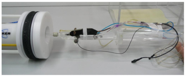
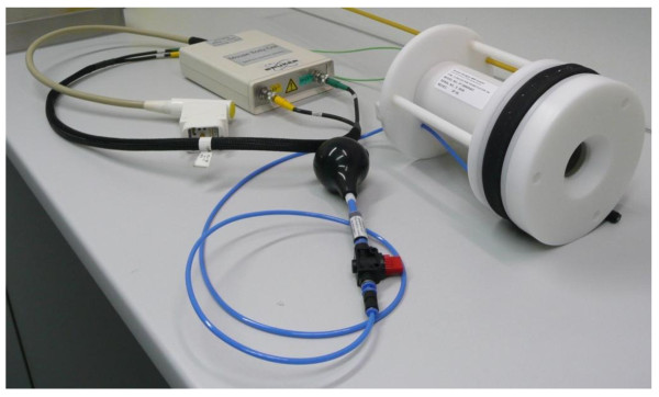
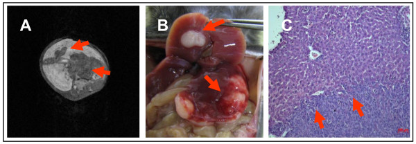
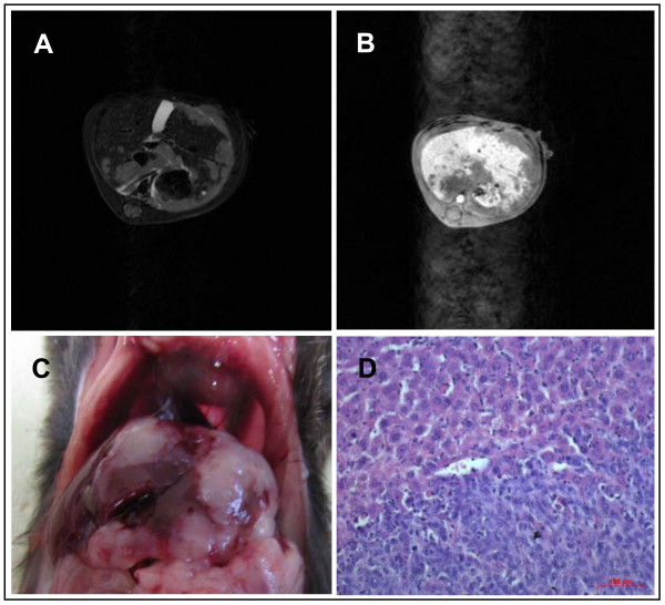
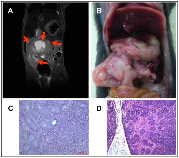
6606PDA murine pancreatic cancer cells were orthotopically injected into the pancreatic head. Liver metastases were induced through splenic injection. Animals were analyzed by MRI three and five weeks following injection. Tumours were detected using T2-weighted high resolution sequences. Tumour volumes were determined by callipers and MRI. Liver metastases were analyzed using gadolinium-EOB-DTPA and T1-weighted 3D-Flash sequences. Tumour blood flow was measured using low molecular gadobutrol and high molecular gadolinium-DTPA.
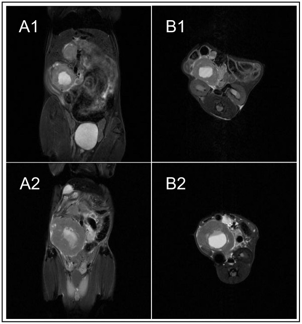
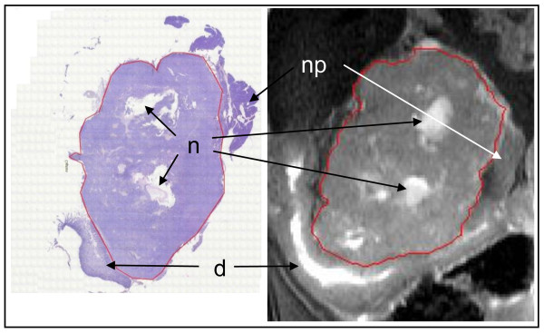
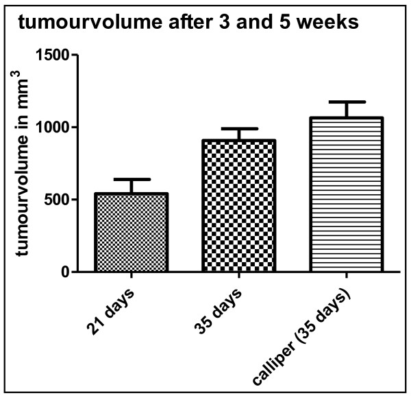
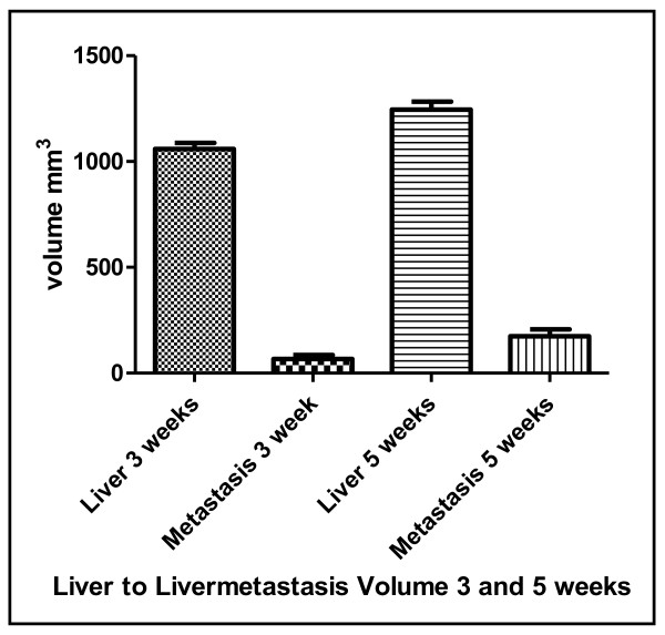
MRI handling and applicability was similar to human systems, resolution as low as 0.1 mm. After 5 weeks tumour volumes differed significantly (p < 0.01) when comparing calliper measurments (n = 5, mean 1065 mm3+/-243 mm3) with MRI (mean 918 mm3+/-193 mm3) with MRI being more precise. Histology (n = 5) confirmed MRI tumour measurements (mean size MRI 38.5 mm2+/-22.8 mm2 versus 32.6 mm2+/-22.6 mm2 (histology), p < 0,0004) with differences due to fixation and processing of specimens. After splenic injection all mice developed liver metastases with a mean of 8 metastases and a mean volume of 173.8 mm3+/-56.7 mm3 after 5 weeks. Lymphnodes were also easily identified. Tumour accumulation of gadobutrol was significantly (p < 0.05) higher than gadolinium-DTPA. All imaging experiments could be done repeatedly to comply with the 3R-principle thus reducing the number of experimental animals.
This model permits monitoring of tumour growth and metastasis formation in longitudinal non-invasive high-resolution MR studies including using contrast agents comparable to human pancreatic cancer. This multidisciplinary environment enables radiologists, surgeons and physicians to further improve translational research and therapies of pancreatic cancer.
胰腺癌是西方世界第四大肿瘤死亡原因。然而,合适的肿瘤模型却很匮乏。在这里,我们提出了一种使用 7 特斯拉 MRI 的同种异体小鼠胰腺癌模型,并评估了其临床相关性和适用性。
将 6606PDA 小鼠胰腺癌细胞原位注射到头胰腺。通过脾内注射诱导肝转移。在注射后 3 周和 5 周时,通过 MRI 对动物进行分析。使用 T2 加权高分辨率序列检测肿瘤。通过卡尺和 MRI 确定肿瘤体积。使用钆塞酸二钠和 T1 加权 3D-Flash 序列分析肝转移。使用低分子钆布醇和高分子钆-DTPA 测量肿瘤血流。
MRI 操作和适用性与人类系统相似,分辨率低至 0.1 毫米。5 周后,卡尺测量的肿瘤体积(n = 5,平均 1065 mm3±243 mm3)与 MRI 测量的肿瘤体积(n = 5,平均 918 mm3±193 mm3)差异有统计学意义(p < 0.01),MRI 更精确。组织学(n = 5)证实了 MRI 肿瘤测量(平均大小 MRI 38.5 mm2±22.8 mm2 与 32.6 mm2±22.6 mm2(组织学),p < 0.0004),差异是由于标本的固定和处理造成的。脾内注射后,所有小鼠均发展为肝转移,5 周后平均有 8 个转移灶,平均体积为 173.8 mm3±56.7 mm3。淋巴结也很容易识别。与钆-DTPA 相比,钆布醇的肿瘤积聚明显更高(p < 0.05)。所有成像实验都可以重复进行,以符合 3R 原则,从而减少实验动物的数量。
该模型允许在纵向非侵入性高分辨率磁共振研究中监测肿瘤生长和转移形成,包括使用与人胰腺癌相似的对比剂。这种多学科环境使放射科医生、外科医生和内科医生能够进一步改进胰腺癌的转化研究和治疗。