• 文献检索
  • 文档翻译
  • 深度研究
  • 学术资讯
  • Suppr Zotero 插件Zotero 插件
  • 邀请有礼
  • 套餐&价格
  • 历史记录
应用&插件
Suppr Zotero 插件Zotero 插件浏览器插件Mac 客户端Windows 客户端微信小程序
定价
高级版会员购买积分包购买API积分包
服务
文献检索文档翻译深度研究API 文档MCP 服务
关于我们
关于 Suppr公司介绍联系我们用户协议隐私条款
关注我们

Suppr 超能文献

核心技术专利:CN118964589B侵权必究
粤ICP备2023148730 号-1Suppr @ 2026

文献检索

告别复杂PubMed语法,用中文像聊天一样搜索,搜遍4000万医学文献。AI智能推荐,让科研检索更轻松。

立即免费搜索

文件翻译

保留排版,准确专业,支持PDF/Word/PPT等文件格式,支持 12+语言互译。

免费翻译文档

深度研究

AI帮你快速写综述,25分钟生成高质量综述,智能提取关键信息,辅助科研写作。

立即免费体验

利用高斯网络模型鉴定蛋白质的配体结合位点。

Identification of ligand binding sites of proteins using the Gaussian Network Model.

机构信息

Center for Computational Biology and Bioinformatics, Koc University, Istanbul Turkey.

出版信息

PLoS One. 2011 Jan 25;6(1):e16474. doi: 10.1371/journal.pone.0016474.

DOI:10.1371/journal.pone.0016474
PMID:21283550
原文链接:https://pmc.ncbi.nlm.nih.gov/articles/PMC3026835/
Abstract

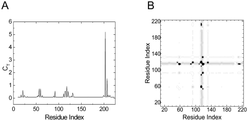
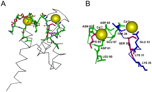
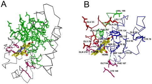
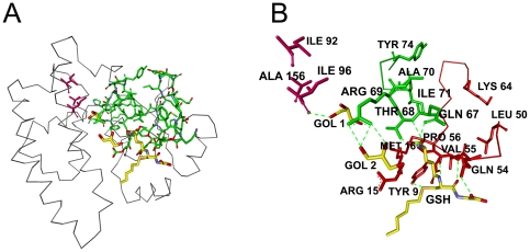
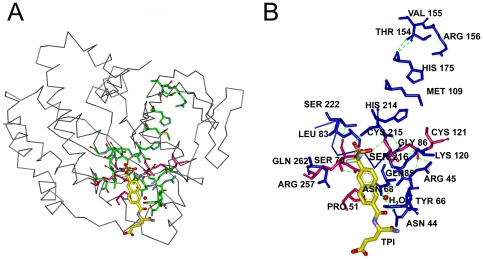
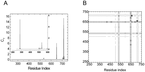
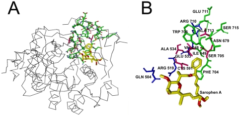
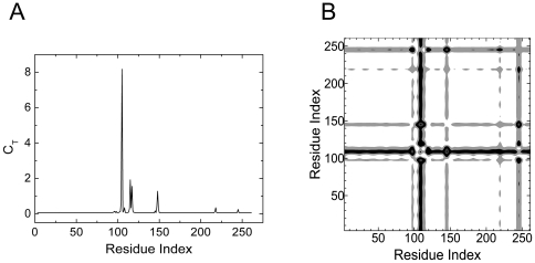
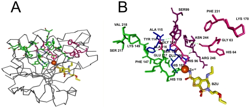
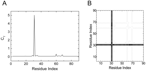
The nonlocal nature of the protein-ligand binding problem is investigated via the Gaussian Network Model with which the residues lying along interaction pathways in a protein and the residues at the binding site are predicted. The predictions of the binding site residues are verified by using several benchmark systems where the topology of the unbound protein and the bound protein-ligand complex are known. Predictions are made on the unbound protein. Agreement of results with the bound complexes indicates that the information for binding resides in the unbound protein. Cliques that consist of three or more residues that are far apart along the primary structure but are in contact in the folded structure are shown to be important determinants of the binding problem. Comparison with known structures shows that the predictive capability of the method is significant.

摘要

通过高斯网络模型研究蛋白质-配体结合问题的非局部性质,该模型可以预测蛋白质中沿相互作用途径的残基和结合部位的残基。通过使用几个已知未结合蛋白质和结合蛋白质-配体复合物拓扑结构的基准系统来验证结合部位残基的预测。在未结合的蛋白质上进行预测。与结合复合物的结果一致表明,结合信息存在于未结合的蛋白质中。沿着一级结构相距很远但在折叠结构中接触的包含三个或更多残基的团簇被证明是结合问题的重要决定因素。与已知结构的比较表明,该方法具有显著的预测能力。

https://cdn.ncbi.nlm.nih.gov/pmc/blobs/c681/3026835/7cbe4c339a61/pone.0016474.g012.jpg
https://cdn.ncbi.nlm.nih.gov/pmc/blobs/c681/3026835/e56051037a7d/pone.0016474.g001.jpg
https://cdn.ncbi.nlm.nih.gov/pmc/blobs/c681/3026835/70a3aad3b95d/pone.0016474.g002.jpg
https://cdn.ncbi.nlm.nih.gov/pmc/blobs/c681/3026835/f607acedca9d/pone.0016474.g003.jpg
https://cdn.ncbi.nlm.nih.gov/pmc/blobs/c681/3026835/5d93d5ac78ff/pone.0016474.g004.jpg
https://cdn.ncbi.nlm.nih.gov/pmc/blobs/c681/3026835/7c822d1b3d23/pone.0016474.g005.jpg
https://cdn.ncbi.nlm.nih.gov/pmc/blobs/c681/3026835/6892fb738248/pone.0016474.g006.jpg
https://cdn.ncbi.nlm.nih.gov/pmc/blobs/c681/3026835/0812bd9febdf/pone.0016474.g007.jpg
https://cdn.ncbi.nlm.nih.gov/pmc/blobs/c681/3026835/a9ca8e43db79/pone.0016474.g008.jpg
https://cdn.ncbi.nlm.nih.gov/pmc/blobs/c681/3026835/30e71370a3bc/pone.0016474.g009.jpg
https://cdn.ncbi.nlm.nih.gov/pmc/blobs/c681/3026835/23cc25ed0f5a/pone.0016474.g010.jpg
https://cdn.ncbi.nlm.nih.gov/pmc/blobs/c681/3026835/2adcdedd22c1/pone.0016474.g011.jpg
https://cdn.ncbi.nlm.nih.gov/pmc/blobs/c681/3026835/7cbe4c339a61/pone.0016474.g012.jpg

相似文献

1
Identification of ligand binding sites of proteins using the Gaussian Network Model.利用高斯网络模型鉴定蛋白质的配体结合位点。
PLoS One. 2011 Jan 25;6(1):e16474. doi: 10.1371/journal.pone.0016474.
2
The roles of contact residue disorder and domain composition in characterizing protein-ligand binding specificity and promiscuity.接触残基无序和结构域组成在表征蛋白质-配体结合特异性和多配体结合性中的作用。
Mol Biosyst. 2011 Dec;7(12):3280-6. doi: 10.1039/c1mb05325f. Epub 2011 Oct 14.
3
Structure-Based Analysis of Cryptic-Site Opening.基于结构的隐匿位点开启分析。
Structure. 2020 Feb 4;28(2):223-235.e2. doi: 10.1016/j.str.2019.11.007. Epub 2019 Dec 3.
4
Motions of Allosteric and Orthosteric Ligand-Binding Sites in Proteins are Highly Correlated.蛋白质中的变构和变构配体结合位点的运动高度相关。
J Chem Inf Model. 2016 Sep 26;56(9):1725-33. doi: 10.1021/acs.jcim.6b00039. Epub 2016 Sep 12.
5
Auto-FACE: an NMR based binding site mapping program for fast chemical exchange protein-ligand systems.Auto-FACE:一种基于 NMR 的结合位点映射程序,用于快速化学交换蛋白-配体体系。
PLoS One. 2010 Feb 18;5(2):e8943. doi: 10.1371/journal.pone.0008943.
6
Protein-protein docking with multiple residue conformations and residue substitutions.具有多个残基构象和残基取代的蛋白质-蛋白质对接
Protein Sci. 2002 Jun;11(6):1393-408. doi: 10.1110/ps.2830102.
7
Prediction of binding sites in receptor-ligand complexes with the Gaussian Network Model.利用高斯网络模型预测受体-配体复合物中的结合位点。
Phys Rev Lett. 2008 Jun 6;100(22):228102. doi: 10.1103/PhysRevLett.100.228102. Epub 2008 Jun 3.
8
An Analysis of Central Residues Between Ligand-Bound and Ligand-Free Protein Structures Based on Network Approach.基于网络方法对配体结合和无配体蛋白质结构之间中心残基的分析
Protein Pept Lett. 2017 Aug;24(6):517-527. doi: 10.2174/0929866524666170413120940.
9
Topological Analyses of Protein-Ligand Binding: a Network Approach.蛋白质-配体结合的拓扑分析:一种网络方法。
Curr Protein Pept Sci. 2016;17(1):37-40. doi: 10.2174/1389203716666150923103934.
10
Identification and Analysis of Key Residues Involved in Folding and Binding of Protein-carbohydrate Complexes.蛋白质-碳水化合物复合物折叠和结合过程中关键残基的鉴定与分析
Protein Pept Lett. 2018;25(4):379-389. doi: 10.2174/0929866525666180221122529.

引用本文的文献

1
Estimating the Similarity between Protein Pockets.估算蛋白质口袋之间的相似性。
Int J Mol Sci. 2022 Oct 18;23(20):12462. doi: 10.3390/ijms232012462.
2
Comparative effects of oncogenic mutations G12C, G12V, G13D, and Q61H on local conformations and dynamics of K-Ras.致癌突变G12C、G12V、G13D和Q61H对K-Ras局部构象和动力学的比较影响。
Comput Struct Biotechnol J. 2020 Apr 9;18:1000-1011. doi: 10.1016/j.csbj.2020.04.003. eCollection 2020.
3
Interpreting the Dynamics of Binding Interactions of snRNA and U1A Using a Coarse-Grained Model.

本文引用的文献

1
Predicting important residues and interaction pathways in proteins using Gaussian Network Model: binding and stability of HLA proteins.使用高斯网络模型预测蛋白质中的重要残基和相互作用途径:HLA 蛋白的结合和稳定性。
PLoS Comput Biol. 2010 Jul 8;6(7):e1000845. doi: 10.1371/journal.pcbi.1000845.
2
Structural and kinetic study of the extended active site for proton transfer in human carbonic anhydrase II.人碳酸酐酶 II 中质子转移的扩展活性位点的结构和动力学研究。
Biochemistry. 2010 Aug 3;49(30):6394-9. doi: 10.1021/bi1007645.
3
Elucidation of the proton transport mechanism in human carbonic anhydrase II.
使用粗粒化模型来解释 snRNA 和 U1A 结合相互作用的动力学。
Biophys J. 2019 May 7;116(9):1625-1636. doi: 10.1016/j.bpj.2019.03.008. Epub 2019 Mar 21.
4
Gaussian network model can be enhanced by combining solvent accessibility in proteins.高斯网络模型可以通过结合蛋白质中的溶剂可及性来增强。
Sci Rep. 2017 Aug 8;7(1):7486. doi: 10.1038/s41598-017-07677-9.
5
Identification of Hot Spots in Protein Structures Using Gaussian Network Model and Gaussian Naive Bayes.使用高斯网络模型和高斯朴素贝叶斯识别蛋白质结构中的热点
Biomed Res Int. 2016;2016:4354901. doi: 10.1155/2016/4354901. Epub 2016 Nov 2.
6
Regulation of ryanodine receptor RyR2 by protein-protein interactions: prediction of a PKA binding site on the N-terminal domain of RyR2 and its relation to disease causing mutations.通过蛋白质-蛋白质相互作用对兰尼碱受体RyR2的调控:预测RyR2 N端结构域上的蛋白激酶A结合位点及其与致病突变的关系
F1000Res. 2015 Jan 28;4:29. doi: 10.12688/f1000research.5858.1. eCollection 2015.
7
Sequence-based Gaussian network model for protein dynamics.基于序列的蛋白质动力学高斯网络模型。
Bioinformatics. 2014 Feb 15;30(4):497-505. doi: 10.1093/bioinformatics/btt716. Epub 2013 Dec 12.
8
A quantitative measure of electrostatic perturbation in holo and apo enzymes induced by structural changes.一种通过结构变化诱导的全酶和脱辅基酶中静电微扰的定量测量方法。
PLoS One. 2013;8(3):e59352. doi: 10.1371/journal.pone.0059352. Epub 2013 Mar 14.
人类碳酸酐酶II中质子转运机制的阐释。
J Am Chem Soc. 2009 Jun 10;131(22):7598-608. doi: 10.1021/ja8091938.
4
Statistical thermodynamics of residue fluctuations in native proteins.天然蛋白质中残基波动的统计热力学
J Chem Phys. 2009 Mar 7;130(9):095103. doi: 10.1063/1.3078517.
5
Analysis of correlations between energy and residue fluctuations in native proteins and determination of specific sites for binding.天然蛋白质中能量与残基波动之间的相关性分析以及结合特异性位点的确定。
Phys Rev Lett. 2009 Feb 27;102(8):088103. doi: 10.1103/PhysRevLett.102.088103.
6
ACC2 is expressed at high levels in human white adipose and has an isoform with a novel N-terminus [corrected].ACC2在人类白色脂肪中高水平表达,并且有一种具有新N端的同工型[已修正]。
PLoS One. 2009;4(2):e4369. doi: 10.1371/journal.pone.0004369. Epub 2009 Feb 3.
7
Prediction of binding sites in receptor-ligand complexes with the Gaussian Network Model.利用高斯网络模型预测受体-配体复合物中的结合位点。
Phys Rev Lett. 2008 Jun 6;100(22):228102. doi: 10.1103/PhysRevLett.100.228102. Epub 2008 Jun 3.
8
A method of matrix analysis of group structure.一种群体结构的矩阵分析方法。
Psychometrika. 1949 Jun;14(2):95-116. doi: 10.1007/BF02289146.
9
Structures and metal-ion-binding properties of the Ca2+-binding helix-loop-helix EF-hand motifs.钙离子结合螺旋-环-螺旋EF手基序的结构与金属离子结合特性
Biochem J. 2007 Jul 15;405(2):199-221. doi: 10.1042/BJ20070255.
10
Protein tyrosine phosphatases: from genes, to function, to disease.蛋白质酪氨酸磷酸酶:从基因到功能再到疾病
Nat Rev Mol Cell Biol. 2006 Nov;7(11):833-46. doi: 10.1038/nrm2039.