Department of Pathology, School of Medicine and Health Sciences, University of North Dakota, Grand Forks, ND, USA.
Cancer Cell Int. 2011 Feb 8;11(1):2. doi: 10.1186/1475-2867-11-2.
Studies have shown that metallothionein 3 (MT-3) is not expressed in normal urothelium or in the UROtsa cell line, but is expressed in urothelial cancer and in tumors generated from the UROtsa cells that have been transformed by cadmium (Cd+2) or arsenite (As+3).The present study had two major goals. One, to determine if epigenetic modifications control urothelial MT-3 gene expression and if regulation is altered by malignant transformation by Cd+2 or As+3. Two, to determine if MT-3 expression might translate clinically as a biomarker for malignant urothelial cells released into the urine.
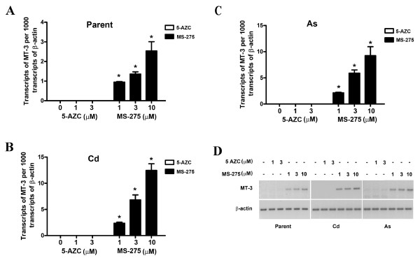
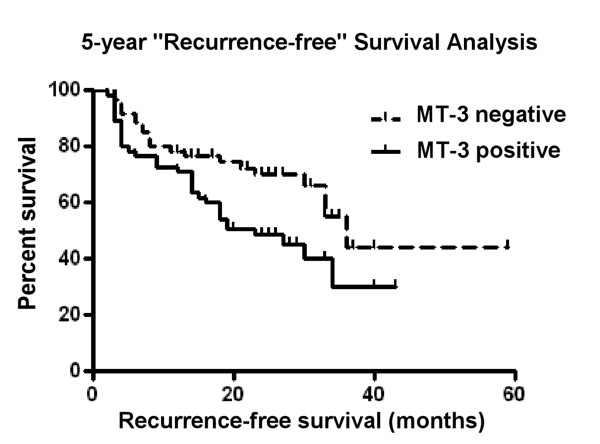
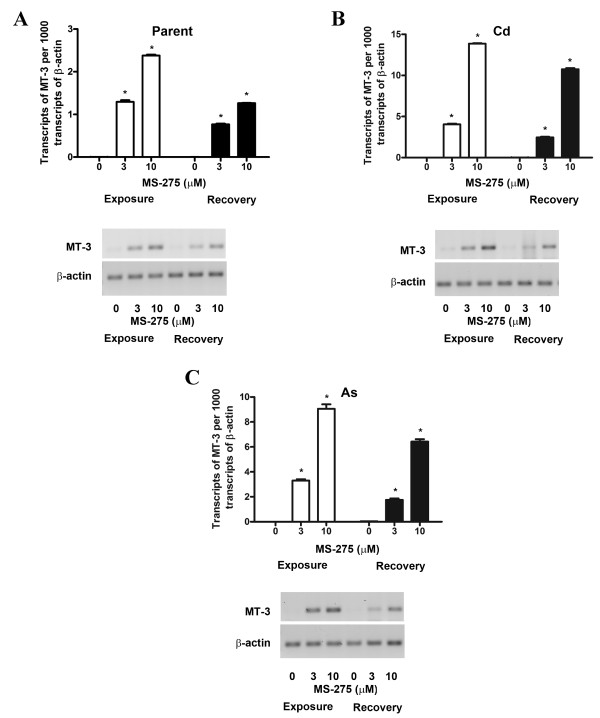
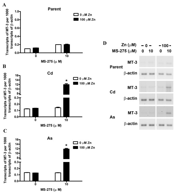
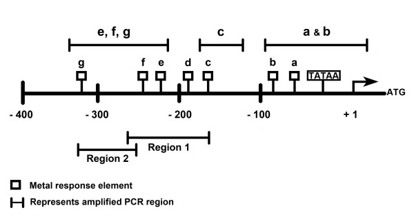
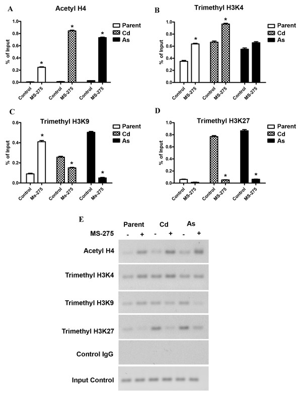
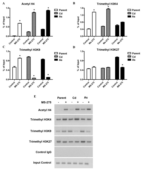
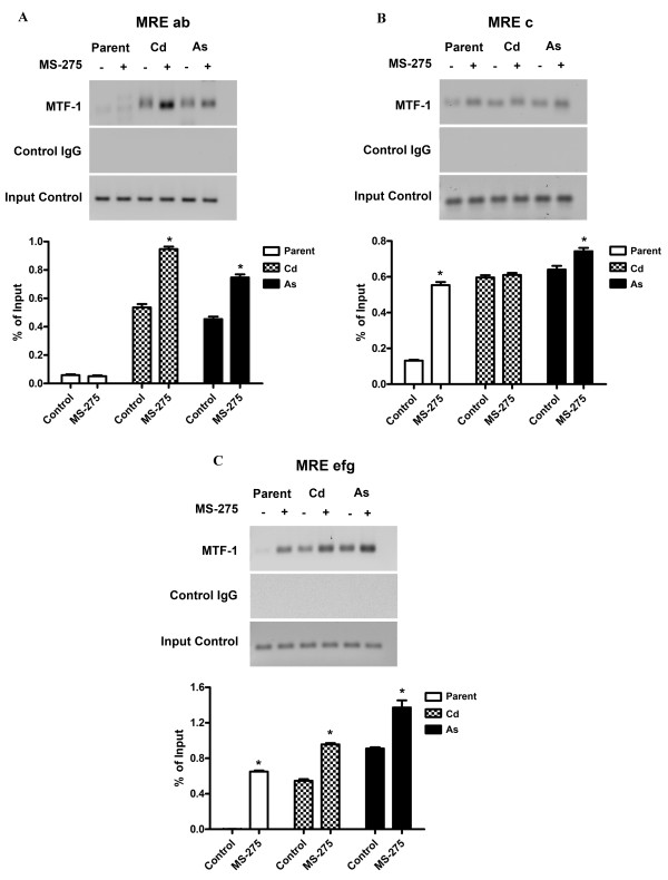
The histone deacetylase inhibitor MS-275 induced MT-3 mRNA expression in both parental UROtsa cells and their transformed counterparts. The demethylating agent, 5-Aza-2'-deoxycytidine (5-AZC) had no effect on MT-3 mRNA expression. ChIP analysis showed that metal-responsive transformation factor-1 (MTF-1) binding to metal response elements (MRE) elements of the MT-3 promoter was restricted in parental UROtsa cells, but MTF-1 binding to the MREs was unrestricted in the transformed cell lines. Histone modifications at acetyl H4, trimethyl H3K4, trimethyl H3K27, and trimethyl H3K9 were compared between the parental and transformed cell lines in the presence and absence of MS-275. The pattern of histone modifications suggested that the MT-3 promoter in the Cd+2 and As+3 transformed cells has gained bivalent chromatin structure, having elements of being "transcriptionally repressed" and "transcription ready", when compared to parental cells. An analysis of MT-3 staining in urinary cytologies showed that a subset of both active and non-active patients with urothelial cancer shed positive cells in their urine, but that control patients only rarely shed MT-3 positive cells.
The MT-3 gene is silenced in non-transformed urothelial cells by a mechanism involving histone modification of the MT-3 promoter. In contrast, transformation of the urothelial cells with either Cd+2 or As+3 modified the chromatin of the MT-3 promoter to a bivalent state of promoter readiness. Urinary cytology for MT-3 positive cells would not improve the diagnosis of urothelial cancer, but might have potential as a biomarker for tumor progression.
研究表明,金属硫蛋白 3(MT-3)在正常尿路上皮或 UROtsa 细胞系中不表达,但在膀胱癌和由镉(Cd+2)或亚砷酸盐(As+3)转化的 UROtsa 细胞生成的肿瘤中表达。本研究有两个主要目标。一是确定表观遗传修饰是否控制尿路上皮 MT-3 基因的表达,以及 Cd+2 或 As+3 引起的恶性转化是否改变了这种调节。二是确定 MT-3 表达是否可能作为释放到尿液中的恶性尿路上皮细胞的临床生物标志物进行翻译。
组蛋白去乙酰化酶抑制剂 MS-275 诱导亲本 UROtsa 细胞及其转化对应物的 MT-3 mRNA 表达。去甲基化剂 5-氮杂-2'-脱氧胞苷(5-AZC)对 MT-3 mRNA 表达没有影响。ChIP 分析表明,金属反应性转化因子-1(MTF-1)与 MT-3 启动子的金属反应元件(MRE)结合在亲本 UROtsa 细胞中受到限制,但在转化细胞系中 MTF-1 与 MRE 的结合不受限制。在存在和不存在 MS-275 的情况下,比较了亲本和转化细胞系之间乙酰化 H4、三甲基 H3K4、三甲基 H3K27 和三甲基 H3K9 组蛋白修饰的模式。与亲本细胞相比,Cd+2 和 As+3 转化细胞中的 MT-3 启动子的组蛋白修饰模式表明,该启动子具有“转录抑制”和“转录准备”的特征,获得了双价染色质结构。尿细胞学中 MT-3 染色的分析表明,患有膀胱癌的活跃和非活跃患者的亚群在尿液中脱落阳性细胞,但对照患者很少脱落 MT-3 阳性细胞。
非转化尿路上皮细胞中的 MT-3 基因通过涉及 MT-3 启动子组蛋白修饰的机制被沉默。相比之下,用 Cd+2 或 As+3 转化尿路上皮细胞会将 MT-3 启动子的染色质修饰为启动子准备的双价状态。尿细胞学中 MT-3 阳性细胞的检测不会提高膀胱癌的诊断,但可能作为肿瘤进展的生物标志物具有潜力。