Centre for Infection and Immunity, School of Medicine, Dentistry and Biomedical Sciences, Queen's University of Belfast, Belfast, United Kingdom.
PLoS Pathog. 2011 Jan 27;7(1):e1001263. doi: 10.1371/journal.ppat.1001263.
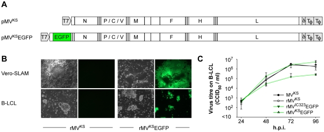
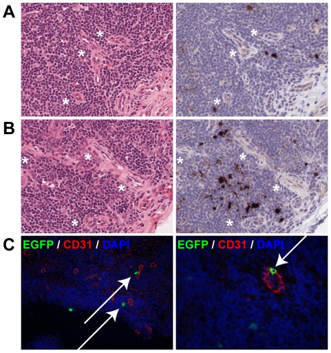
Measles virus (MV) is highly infectious, and has long been thought to enter the host by infecting epithelial cells of the respiratory tract. However, epithelial cells do not express signaling lymphocyte activation molecule (CD150), which is the high-affinity cellular receptor for wild-type MV strains. We have generated a new recombinant MV strain expressing enhanced green fluorescent protein (EGFP), based on a wild-type genotype B3 virus isolate from Khartoum, Sudan (KS). Cynomolgus macaques were infected with a high dose of rMV(KS)EGFP by aerosol inhalation to ensure that the virus could reach the full range of potential target cells throughout the entire respiratory tract. Animals were euthanized 2, 3, 4 or 5 days post-infection (d.p.i., n = 3 per time point) and infected (EGFP(+)) cells were identified at all four time points, albeit at low levels 2 and 3 d.p.i. At these earliest time points, MV-infected cells were exclusively detected in the lungs by fluorescence microscopy, histopathology and/or virus isolation from broncho-alveolar lavage cells. On 2 d.p.i., EGFP(+) cells were phenotypically typed as large mononuclear cells present in the alveolar lumen or lining the alveolar epithelium. One to two days later, larger clusters of MV-infected cells were detected in bronchus-associated lymphoid tissue (BALT) and in the tracheo-bronchial lymph nodes. From 4 d.p.i. onward, MV-infected cells were detected in peripheral blood and various lymphoid tissues. In spite of the possibility for the aerosolized virus to infect cells and lymphoid tissues of the upper respiratory tract, MV-infected cells were not detected in either the tonsils or the adenoids until after onset of viremia. These data strongly suggest that in our model MV entered the host at the alveolar level by infecting macrophages or dendritic cells, which traffic the virus to BALT or regional lymph nodes, resulting in local amplification and subsequent systemic dissemination by viremia.
麻疹病毒(MV)具有高度传染性,长期以来被认为通过感染呼吸道的上皮细胞进入宿主。然而,上皮细胞不表达信号淋巴细胞激活分子(CD150),这是野生型 MV 株的高亲和力细胞受体。我们基于来自苏丹喀土穆的 B3 病毒分离株,生成了一种表达增强型绿色荧光蛋白(EGFP)的新型重组 MV 株(rMV(KS)EGFP)。恒河猴通过气溶胶吸入感染高剂量 rMV(KS)EGFP,以确保病毒能够到达整个呼吸道的全部潜在靶细胞。动物在感染后 2、3、4 或 5 天(n=每个时间点 3 只)安乐死,并在所有四个时间点均鉴定出感染(EGFP(+))细胞,尽管在 2 和 3 天感染时水平较低。在这些最早的时间点,通过荧光显微镜、组织病理学和/或从支气管肺泡灌洗细胞中分离病毒,仅在肺部检测到 MV 感染的细胞。在 2 天感染时,EGFP(+)细胞被表型鉴定为存在于肺泡腔中的大单核细胞或沿肺泡上皮排列。一到两天后,在支气管相关淋巴组织(BALT)和气管支气管淋巴结中检测到更大的 MV 感染细胞簇。从 4 天感染时开始,MV 感染的细胞在外周血和各种淋巴组织中被检测到。尽管雾化病毒有可能感染上呼吸道的细胞和淋巴组织,但直到病毒血症开始后,才在扁桃体或腺样体中检测到 MV 感染的细胞。这些数据强烈表明,在我们的模型中,MV 通过感染巨噬细胞或树突状细胞进入肺泡水平的宿主,这些细胞将病毒运送到 BALT 或局部淋巴结,导致局部扩增,随后通过病毒血症进行系统传播。