Shetty Devi Charan, Ahuja Puneet, Taneja D K, Rathore Ajit Singh, Chhina Shivjot, Ahuja Upasana Sethi, Kumar Kiran, Ahuja Anshuman, Rastogi Priyanka
Department of Oral & Maxillofacial Pathology, I.T.S-CDSR, Muradnagar, Ghaziabad, Uttar Pradesh, India.
Vasc Health Risk Manag. 2011 Jan 27;7:41-7. doi: 10.2147/VHRM.S15384.
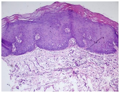
Tumor angiogenesis occurs by recruitment of endothelial cell precursors or by sprouting of existing capillaries, which differ from the normal vasculature by having an altered morphology that can be exploited for diagnosis and as a prognostic indicator. Improved technologies have propelled diagnosis into a new era. These technologies have to be used with great precision. The diagnosis of a dysplastic premalignant lesion of the oral mucosa cannot be based solely on clinical findings. Therefore histologic evaluation of a representative biopsy specimen is necessary. Accurate judgment of the proper site for biopsy is essential for reaching a correct diagnosis. The aim of this report is to analyze the vascular patterns with the help of direct oral microscopy and the technique of stereo-optical microscopy in the oral cavity to select biopsy sites, and compare the outcome of a directed biopsy with that of biopsy specimens obtained from sites selected solely on the basis of clinical criteria. The study sample comprised 50 oral mucosal lesions. A statistically significant difference was noted between samples judged to be microscopically representative sites. We conclude that this method would aid in early and better diagnosis and treatment planning of oral premalignant and malignant lesions by assessing the various vascular patterns in the mucosa.
肿瘤血管生成通过募集内皮细胞前体或现有毛细血管的芽生而发生,其与正常脉管系统的不同之处在于具有可用于诊断和作为预后指标的改变的形态。改进的技术已将诊断推进到一个新时代。这些技术必须精确使用。口腔黏膜发育异常的癌前病变诊断不能仅基于临床发现。因此,对代表性活检标本进行组织学评估是必要的。准确判断活检的合适部位对于做出正确诊断至关重要。本报告的目的是借助口腔直接显微镜检查和立体光学显微镜技术分析口腔内的血管模式,以选择活检部位,并将定向活检的结果与仅根据临床标准选择部位获得的活检标本的结果进行比较。研究样本包括50个口腔黏膜病变。在判断为显微镜下代表性部位的样本之间存在统计学上的显著差异。我们得出结论,该方法将通过评估黏膜中的各种血管模式,有助于口腔癌前病变和恶性病变的早期和更好的诊断及治疗规划。