Department of Biomedical Engineering, Cullen College of Engineering, University of Houston, Houston, TX 77204, USA.
J Neuroeng Rehabil. 2011 Feb 27;8:13. doi: 10.1186/1743-0003-8-13.
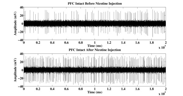
The dopaminergic (DA) neurons in the ventral tegmental area (VTA) are widely implicated in the addiction and natural reward circuitry of the brain. These neurons project to several areas of the brain, including prefrontal cortex (PFC), nucleus accubens (NAc) and amygdala. The functional coupling between PFC and VTA has been demonstrated, but little is known about how PFC mediates nicotinic modulation in VTA DA neurons. The objectives of this study were to investigate the effect of acute nicotine exposure on the VTA DA neuronal firing and to understand how the disruption of communication from PFC affects the firing patterns of VTA DA neurons.
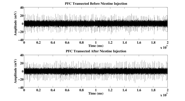
Extracellular single-unit recordings were performed on Sprague-Dawley rats and nicotine was administered after stable recording was established as baseline. In order to test how input from PFC affects the VTA DA neuronal firing, bilateral transections were made immediate caudal to PFC to mechanically delete the interaction between VTA and PFC.
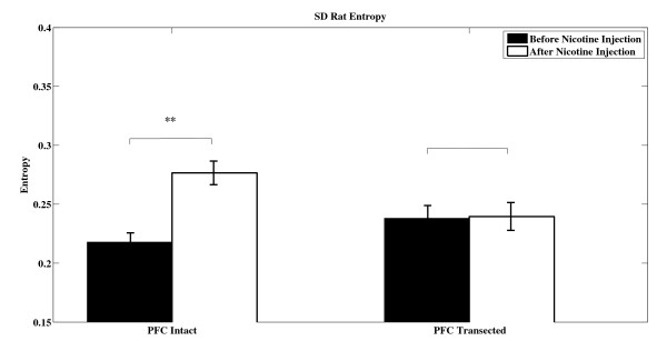
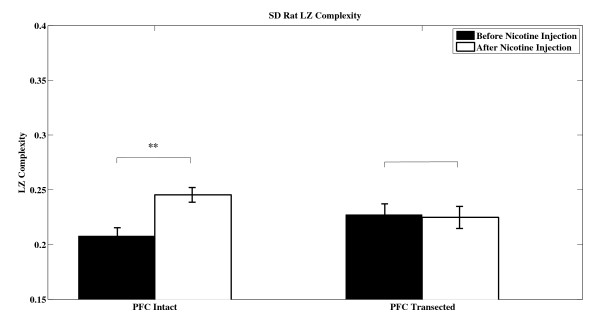
The complexity of the recorded neural firing was subsequently assessed using a method based on the Lempel-Ziv estimator. The results were compared with those obtained when computing the entropy of neural firing. Exposure to nicotine triggered a significant increase in VTA DA neurons firing complexity when communication between PFC and VTA was present, while transection obliterated the effect of nicotine. Similar results were obtained when entropy values were estimated.
Our findings suggest that PFC plays a vital role in mediating VTA activity. We speculate that increased firing complexity with acute nicotine administration in PFC intact subjects is due to the close functional coupling between PFC and VTA. This hypothesis is supported by the fact that deletion of PFC results in minor alterations of VTA DA neural firing when nicotine is acutely administered.
腹侧被盖区(VTA)中的多巴胺能(DA)神经元广泛涉及大脑的成瘾和自然奖励回路。这些神经元投射到大脑的几个区域,包括前额叶皮层(PFC)、伏隔核(NAc)和杏仁核。已经证明了 PFC 和 VTA 之间的功能耦合,但对于 PFC 如何介导 VTA DA 神经元中的烟碱调制知之甚少。本研究的目的是研究急性尼古丁暴露对 VTA DA 神经元放电的影响,并了解 PFC 中断通讯如何影响 VTA DA 神经元的放电模式。
对 Sprague-Dawley 大鼠进行细胞外单单元记录,并在稳定记录建立为基线后给予尼古丁。为了测试 PFC 的输入如何影响 VTA DA 神经元的放电,在 PFC 尾部立即进行双侧横切,以机械消除 VTA 和 PFC 之间的相互作用。
随后使用基于 Lempel-Ziv 估计器的方法评估记录的神经放电的复杂性。将结果与计算神经放电熵时获得的结果进行比较。当 PFC 和 VTA 之间存在通讯时,尼古丁暴露会引发 VTA DA 神经元放电复杂性的显著增加,而横切则消除了尼古丁的作用。当估计熵值时,也得到了类似的结果。
我们的研究结果表明,PFC 在介导 VTA 活动方面起着至关重要的作用。我们推测,在 PFC 完整的受试者中,急性尼古丁给药引起的放电复杂性增加是由于 PFC 和 VTA 之间的紧密功能耦合所致。这一假设得到了以下事实的支持:当急性给予尼古丁时,PFC 的缺失导致 VTA DA 神经元放电的微小改变。