• 文献检索
  • 文档翻译
  • 深度研究
  • 学术资讯
  • Suppr Zotero 插件Zotero 插件
  • 邀请有礼
  • 套餐&价格
  • 历史记录
应用&插件
Suppr Zotero 插件Zotero 插件浏览器插件Mac 客户端Windows 客户端微信小程序
定价
高级版会员购买积分包购买API积分包
服务
文献检索文档翻译深度研究API 文档MCP 服务
关于我们
关于 Suppr公司介绍联系我们用户协议隐私条款
关注我们

Suppr 超能文献

核心技术专利:CN118964589B侵权必究
粤ICP备2023148730 号-1Suppr @ 2026

文献检索

告别复杂PubMed语法,用中文像聊天一样搜索,搜遍4000万医学文献。AI智能推荐,让科研检索更轻松。

立即免费搜索

文件翻译

保留排版,准确专业,支持PDF/Word/PPT等文件格式,支持 12+语言互译。

免费翻译文档

深度研究

AI帮你快速写综述,25分钟生成高质量综述,智能提取关键信息,辅助科研写作。

立即免费体验

相似文献

1
Dynamic imaging of ictal oscillations using non-invasive high-resolution EEG.使用非侵入性高分辨率 EEG 进行癫痫发作期振荡的动态成像。
Neuroimage. 2011 Jun 15;56(4):1908-17. doi: 10.1016/j.neuroimage.2011.03.043. Epub 2011 Mar 29.
2
EEG source imaging of epileptic activity at seizure onset.癫痫发作起始时的癫痫活动的脑电图源成像。
Epilepsy Res. 2018 Oct;146:160-171. doi: 10.1016/j.eplepsyres.2018.07.006. Epub 2018 Jul 27.
3
Dynamic imaging of ictal rhythmic activity using dense-array EEG.
Annu Int Conf IEEE Eng Med Biol Soc. 2011;2011:8271-4. doi: 10.1109/IEMBS.2011.6092039.
4
Performance analysis of EEG seizure detection features.脑电癫痫发作检测特征的性能分析。
Epilepsy Res. 2020 Nov;167:106483. doi: 10.1016/j.eplepsyres.2020.106483. Epub 2020 Oct 6.
5
Dynamic imaging of seizure activity in pediatric epilepsy patients.儿童癫痫患者的癫痫活动动态成像。
Clin Neurophysiol. 2012 Nov;123(11):2122-9. doi: 10.1016/j.clinph.2012.04.021. Epub 2012 May 18.
6
Contribution of Ictal Source Imaging for Localizing Seizure Onset Zone in Patients With Focal Epilepsy.癫痫灶定位中发作期源成像的作用。
Neurology. 2021 Jan 19;96(3):e366-e375. doi: 10.1212/WNL.0000000000011109. Epub 2020 Oct 23.
7
Simultaneous scalp EEG improves seizure lateralization during unilateral intracranial EEG evaluation in temporal lobe epilepsy.头皮 EEG 同步监测有助于提高单侧颅内 EEG 评估颞叶癫痫时的癫痫灶侧别定位。
Seizure. 2019 Jan;64:8-15. doi: 10.1016/j.seizure.2018.11.015. Epub 2018 Nov 24.
8
Source localization of the seizure onset zone from ictal EEG/MEG data.基于发作期脑电图/脑磁图数据的癫痫发作起始区的源定位
Hum Brain Mapp. 2016 Jul;37(7):2528-46. doi: 10.1002/hbm.23191. Epub 2016 Apr 5.
9
Ictal high-frequency oscillations in neocortical epilepsy: implications for seizure localization and surgical resection.新皮层癫痫中的发作高频振荡:对发作定位和手术切除的影响。
Epilepsia. 2011 Oct;52(10):1792-801. doi: 10.1111/j.1528-1167.2011.03165.x. Epub 2011 Jul 18.
10
EEG source connectivity to localize the seizure onset zone in patients with drug resistant epilepsy.脑电图源连接以定位耐药性癫痫患者的发作起始区。
Neuroimage Clin. 2017 Sep 14;16:689-698. doi: 10.1016/j.nicl.2017.09.011. eCollection 2017.

引用本文的文献

1
Non-Invasive Localization of Epileptogenic Zone in Drug-Resistant Epilepsy Based on Time-Frequency Analysis and VGG Convolutional Neural Network.基于时频分析和VGG卷积神经网络的耐药性癫痫致痫区无创定位
Bioengineering (Basel). 2025 Apr 23;12(5):443. doi: 10.3390/bioengineering12050443.
2
Seizure Sources Can Be Imaged from Scalp EEG by Means of Biophysically Constrained Deep Neural Networks.癫痫发作源可通过生物物理约束深度神经网络从头皮脑电图成像。
Adv Sci (Weinh). 2024 Dec;11(47):e2405246. doi: 10.1002/advs.202405246. Epub 2024 Oct 29.
3
Electroencephalographic source imaging of spikes with concurrent high-frequency oscillations is concordant with the clinical ground truth.伴有同步高频振荡的棘波的脑电图源成像与临床实际情况相符。
Epilepsia. 2024 Dec;65(12):3571-3582. doi: 10.1111/epi.18141. Epub 2024 Oct 10.
4
Viscous dynamics associated with hypoexcitation and structural disintegration in neurodegeneration via generative whole-brain modeling.通过生成式全脑建模研究神经退行性变中低兴奋性和结构解体相关的粘性动力学。
Alzheimers Dement. 2024 May;20(5):3228-3250. doi: 10.1002/alz.13788. Epub 2024 Mar 19.
5
Combining independent component analysis and source localization for improving spatial sampling of stereoelectroencephalography in epilepsy.结合独立成分分析和源定位以改善癫痫立体脑电图的空间采样
Sci Rep. 2024 Feb 19;14(1):4071. doi: 10.1038/s41598-024-54359-4.
6
Disentanglement of Resting State Brain Networks for Localizing Epileptogenic Zone in Focal Epilepsy.分离静息态脑网络以定位局灶性癫痫的致痫区。
Brain Topogr. 2024 Jan;37(1):152-168. doi: 10.1007/s10548-023-01025-z. Epub 2023 Dec 19.
7
Ictal source localization from intracranial recordings.基于颅内记录的发作期源定位
Clin Neurophysiol. 2022 Dec;144:121-122. doi: 10.1016/j.clinph.2022.09.013. Epub 2022 Oct 6.
8
Deep neural networks constrained by neural mass models improve electrophysiological source imaging of spatiotemporal brain dynamics.受神经质量模型约束的深度神经网络可提高时空脑动力学的电生理源成像。
Proc Natl Acad Sci U S A. 2022 Aug 2;119(31):e2201128119. doi: 10.1073/pnas.2201128119. Epub 2022 Jul 26.
9
Imaging the extent and location of spatiotemporally distributed epileptiform sources from MEG measurements.从脑磁图测量中对时空分布的癫痫源的范围和位置进行成像。
Neuroimage Clin. 2022;33:102903. doi: 10.1016/j.nicl.2021.102903. Epub 2021 Nov 29.
10
Robust EEG Based Biomarkers to Detect Alzheimer's Disease.基于稳健脑电图的阿尔茨海默病检测生物标志物
Brain Sci. 2021 Jul 31;11(8):1026. doi: 10.3390/brainsci11081026.

本文引用的文献

1
EEG-fMRI reciprocal functional neuroimaging.脑电-功能磁共振成像的互功能神经影像学。
Clin Neurophysiol. 2010 Aug;121(8):1240-50. doi: 10.1016/j.clinph.2010.02.153. Epub 2010 Apr 8.
2
Source localization of ictal epileptic activity investigated by high resolution EEG and validated by SEEG.高分辨率 EEG 研究和 SEEG 验证的发作期癫痫活动的源定位。
Neuroimage. 2010 Jun;51(2):642-53. doi: 10.1016/j.neuroimage.2010.02.067. Epub 2010 Mar 4.
3
Comparing noninvasive dense array and intracranial electroencephalography for localization of seizures.比较无创密集阵和颅内脑电图在癫痫定位中的应用。
Neurosurgery. 2010 Feb;66(2):354-62. doi: 10.1227/01.NEU.0000363721.06177.07.
4
Interictal EEG spikes identify the region of electrographic seizure onset in some, but not all, pediatric epilepsy patients.间期 EEG 棘波在一些但不是所有儿科癫痫患者中可识别出电临床发作起始的区域。
Epilepsia. 2010 Apr;51(4):592-601. doi: 10.1111/j.1528-1167.2009.02306.x. Epub 2009 Sep 22.
5
Feasibility of simultaneous intracranial EEG-fMRI in humans: a safety study.在人类中同时进行颅内 EEG-fMRI 的可行性:一项安全性研究。
Neuroimage. 2010 Jan 1;49(1):379-90. doi: 10.1016/j.neuroimage.2009.07.062. Epub 2009 Aug 3.
6
Effect of epilepsy magnetic source imaging on intracranial electrode placement.癫痫磁源成像对颅内电极放置的影响。
Ann Neurol. 2009 Jun;65(6):716-23. doi: 10.1002/ana.21660.
7
Cortical and subcortical networks in human secondarily generalized tonic-clonic seizures.人类继发性全身强直-阵挛性癫痫中的皮质和皮质下网络。
Brain. 2009 Apr;132(Pt 4):999-1012. doi: 10.1093/brain/awp028. Epub 2009 Apr 1.
8
Combined use of non-invasive techniques for improved functional localization for a selected group of epilepsy surgery candidates.联合使用非侵入性技术,以改善选定的一组癫痫手术候选者的功能定位。
Neuroimage. 2009 Apr 1;45(2):342-8. doi: 10.1016/j.neuroimage.2008.12.026. Epub 2008 Dec 30.
9
Independent component analysis of subdurally recorded occipital seizures.
Clin Neurophysiol. 2008 Nov;119(11):2437-46. doi: 10.1016/j.clinph.2008.07.276. Epub 2008 Sep 21.
10
ECoG gamma activity during a language task: differentiating expressive and receptive speech areas.语言任务期间的脑电皮层γ活动:区分表达性和接受性语言区域。
Brain. 2008 Aug;131(Pt 8):2013-27. doi: 10.1093/brain/awn147. Epub 2008 Jul 11.

使用非侵入性高分辨率 EEG 进行癫痫发作期振荡的动态成像。

Dynamic imaging of ictal oscillations using non-invasive high-resolution EEG.

机构信息

Department of Biomedical Engineering, University of Minnesota, Minneapolis, MN, USA.

出版信息

Neuroimage. 2011 Jun 15;56(4):1908-17. doi: 10.1016/j.neuroimage.2011.03.043. Epub 2011 Mar 29.

DOI:10.1016/j.neuroimage.2011.03.043
PMID:21453776
原文链接:https://pmc.ncbi.nlm.nih.gov/articles/PMC3359824/
Abstract

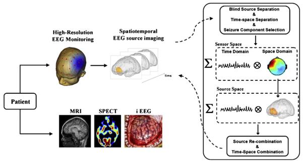
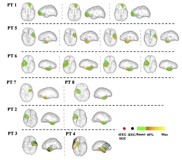
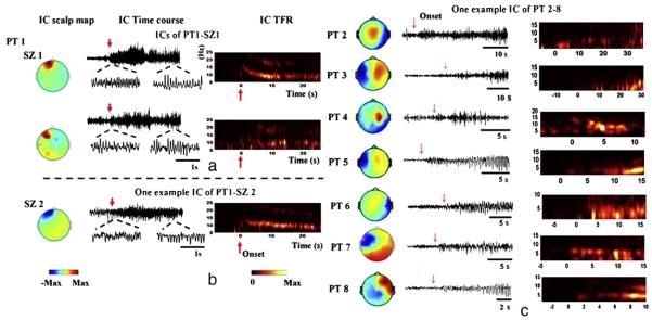
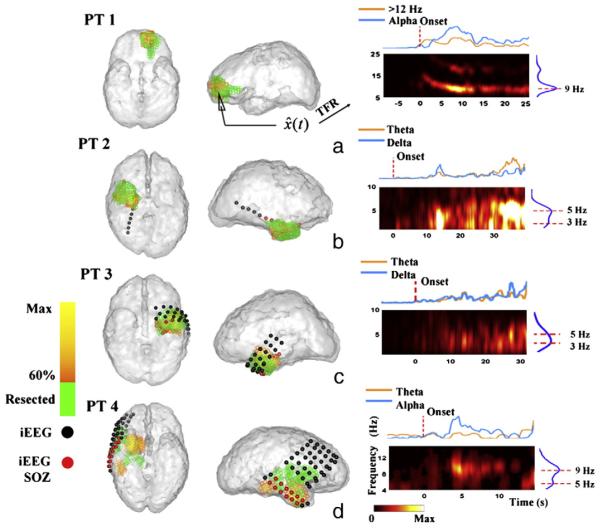
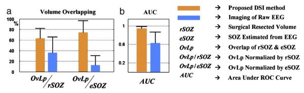
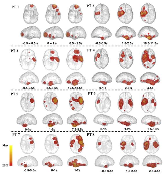
Scalp electroencephalography (EEG) has been established as a major component of the pre-surgical evaluation for epilepsy surgery. However, its ability to localize seizure onset zones (SOZ) has been significantly restricted by its low spatial resolution and indirect correlation with underlying brain activities. Here we report a novel non-invasive dynamic seizure imaging (DSI) approach based upon high-density EEG recordings. This novel approach was particularly designed to image the dynamic changes of ictal rhythmic discharges that evolve through time, space and frequency. This method was evaluated in a group of 8 epilepsy patients and results were rigorously validated using intracranial EEG (iEEG) (n=3) and surgical outcome (n=7). The DSI localized the ictal activity in concordance with surgically resected zones and ictal iEEG recordings in the cohort of patients. The present promising results support the ability to precisely and accurately image dynamic seizure activity from non-invasive measurements. The successful establishment of such a non-invasive seizure imaging modality for surgical evaluation will have a significant impact in the management of medically intractable epilepsy.

摘要

头皮脑电图 (EEG) 已被确立为癫痫手术术前评估的主要组成部分。然而,由于其空间分辨率低且与大脑活动的间接相关性,其定位发作起始区 (SOZ) 的能力受到了很大限制。在这里,我们报告了一种基于高密度 EEG 记录的新型非侵入性动态发作成像 (DSI) 方法。这种新方法是专门设计用来对通过时间、空间和频率演变的发作性节律性放电的动态变化进行成像的。该方法在 8 例癫痫患者中进行了评估,并使用颅内 EEG (iEEG) (n=3) 和手术结果 (n=7) 进行了严格验证。DSI 与患者组中手术切除区域和发作性 iEEG 记录一致地定位了发作活动。目前有希望的结果支持从非侵入性测量中精确和准确地成像动态发作活动的能力。成功建立这种用于手术评估的非侵入性癫痫成像模式将对药物难治性癫痫的治疗产生重大影响。

https://cdn.ncbi.nlm.nih.gov/pmc/blobs/58d4/3359824/c72220708db3/nihms-285693-f0006.jpg
https://cdn.ncbi.nlm.nih.gov/pmc/blobs/58d4/3359824/613904e9d8f5/nihms-285693-f0001.jpg
https://cdn.ncbi.nlm.nih.gov/pmc/blobs/58d4/3359824/ddb97db00814/nihms-285693-f0002.jpg
https://cdn.ncbi.nlm.nih.gov/pmc/blobs/58d4/3359824/6b804732069a/nihms-285693-f0003.jpg
https://cdn.ncbi.nlm.nih.gov/pmc/blobs/58d4/3359824/f2db1680f7c6/nihms-285693-f0004.jpg
https://cdn.ncbi.nlm.nih.gov/pmc/blobs/58d4/3359824/a3e6129e213d/nihms-285693-f0005.jpg
https://cdn.ncbi.nlm.nih.gov/pmc/blobs/58d4/3359824/c72220708db3/nihms-285693-f0006.jpg