College of Life Sciences, Welcome Trust Building, University of Dundee, Dow St., Dundee, UK.
Dev Biol. 2011 Jun 1;354(1):77-86. doi: 10.1016/j.ydbio.2011.03.024. Epub 2011 Mar 31.
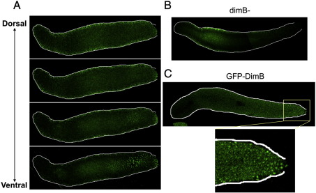
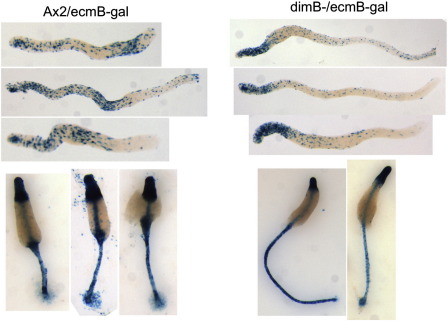
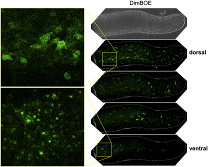
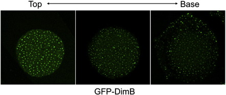
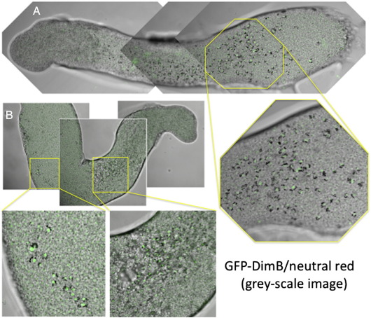
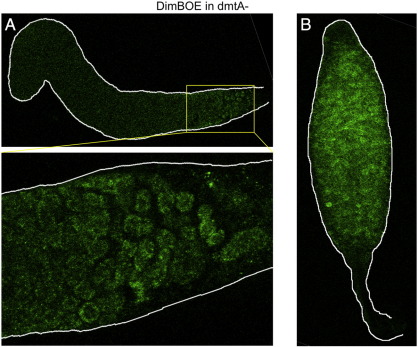
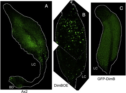
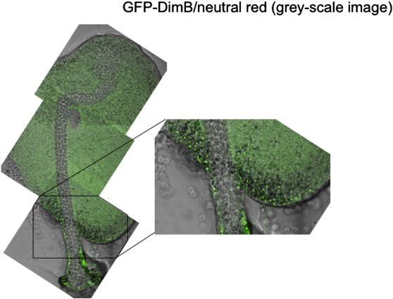
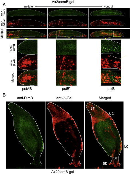
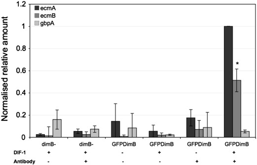
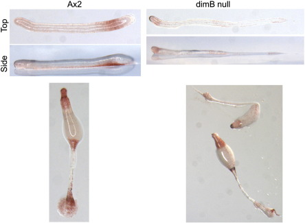
Exposure of monolayer Dictyostelium cells to the signalling polyketide DIF-1 causes DimB, a bZIPtranscription factor, to accumulate in the nucleus where it induces prestalk gene expression. Here we analyse DimB signalling during normal development. In slugs DimB is specifically nuclear enriched in the pstB cells; a cluster of vital dye-staining cells located on the ventral surface of the posterior, prespore region. PstB cells move at culmination, to form the lower cup and the outer basal disc of the fruiting body, and DimB retains a high nuclear concentration in both these tissues. In a dimB null (dimB-) strain there are very few pstB or lower cup cells, as detected by neutral red staining, and it is known that the outer basal disc is absent or much reduced. In the dimB- strain ecmB, a marker of pstB differentiation, is not DIF inducible. Furthermore, ChIP analysis shows that DimB binds to the ecmB promoter in DIF-induced cells. These results suggest that the differentiation of pstB cells is caused by a high perceived level of DIF-1 signalling, leading to nuclear localization of DimB and direct activation of cell type-specific gene expression.
单层盘基网柄菌细胞暴露于信号多酮 DIF-1 会导致 bZIP 转录因子 DimB 积累在核内,从而诱导前柄基因的表达。在这里,我们分析了正常发育过程中的 DimB 信号。在扭结阶段,pstB 细胞中 DimB 特异性地富含于细胞核内;pstB 细胞位于后孢子区腹侧表面的一群重要的染色细胞簇。pstB 细胞在 culmination 时移动,形成子实体的下部杯状结构和外基盘,而 DimB 在这两种组织中都保持着高核浓度。在 dimB 缺失(dimB-)突变体中,通过中性红染色检测到很少有 pstB 或下部杯状细胞,并且已知外基盘缺失或大大减少。在 dimB-突变体 ecmB 中,pstB 分化的标志物,不能被 DIF 诱导。此外,ChIP 分析表明,DimB 在 DIF 诱导的细胞中结合到 ecmB 启动子上。这些结果表明,pstB 细胞的分化是由感知到的高水平 DIF-1 信号引起的,导致 DimB 的核定位和细胞类型特异性基因表达的直接激活。