• 文献检索
  • 文档翻译
  • 深度研究
  • 学术资讯
  • Suppr Zotero 插件Zotero 插件
  • 邀请有礼
  • 套餐&价格
  • 历史记录
应用&插件
Suppr Zotero 插件Zotero 插件浏览器插件Mac 客户端Windows 客户端微信小程序
定价
高级版会员购买积分包购买API积分包
服务
文献检索文档翻译深度研究API 文档MCP 服务
关于我们
关于 Suppr公司介绍联系我们用户协议隐私条款
关注我们

Suppr 超能文献

核心技术专利:CN118964589B侵权必究
粤ICP备2023148730 号-1Suppr @ 2026

文献检索

告别复杂PubMed语法,用中文像聊天一样搜索,搜遍4000万医学文献。AI智能推荐,让科研检索更轻松。

立即免费搜索

文件翻译

保留排版,准确专业,支持PDF/Word/PPT等文件格式,支持 12+语言互译。

免费翻译文档

深度研究

AI帮你快速写综述,25分钟生成高质量综述,智能提取关键信息,辅助科研写作。

立即免费体验

一些荧光蛋白的秘密:在不同的固定液中有明显的漂白现象,以及在 YFP 激发下青色荧光蛋白的光激活。

Some secrets of fluorescent proteins: distinct bleaching in various mounting fluids and photoactivation of cyan fluorescent proteins at YFP-excitation.

机构信息

Department of Vascular Biology and Thrombosis Research, Center for Physiology and Pharmacology, Medical University Vienna, Vienna, Austria.

出版信息

PLoS One. 2011 Apr 7;6(4):e18586. doi: 10.1371/journal.pone.0018586.

DOI:10.1371/journal.pone.0018586
PMID:21490932
原文链接:https://pmc.ncbi.nlm.nih.gov/articles/PMC3072413/
Abstract

BACKGROUND

The use of spectrally distinct variants of green fluorescent protein (GFP) such as cyan or yellow mutants (CFP and YFP, respectively) is very common in all different fields of life sciences, e.g. for marking specific proteins or cells or to determine protein interactions. In the latter case, the quantum physical phenomenon of fluorescence resonance energy transfer (FRET) is exploited by specific microscopy techniques to visualize proximity of proteins.

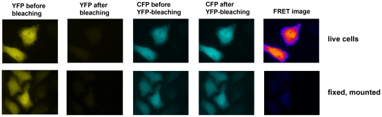
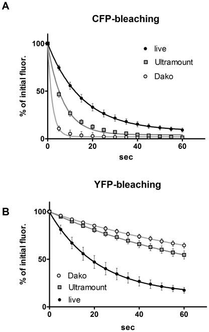
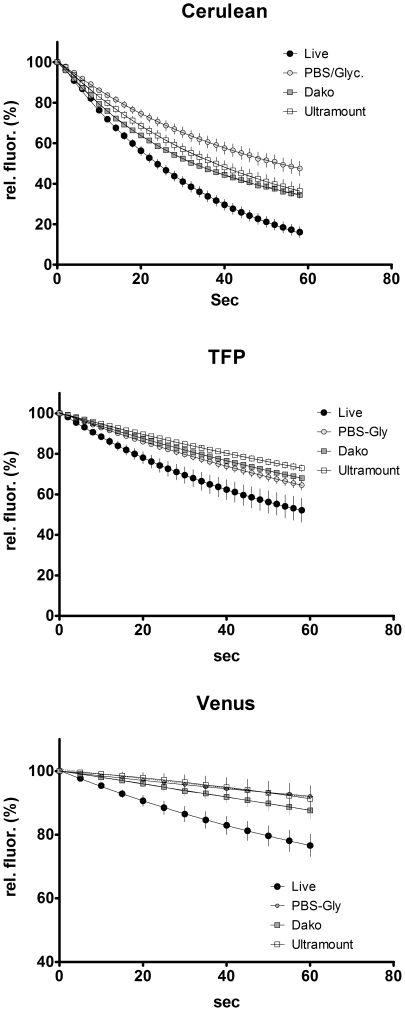
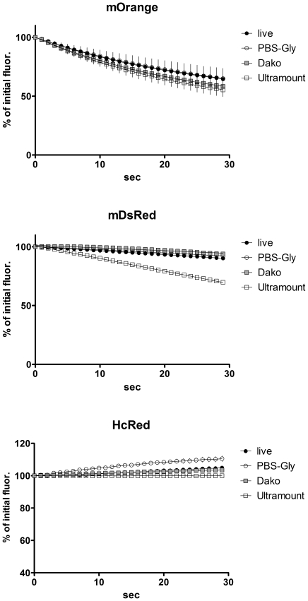
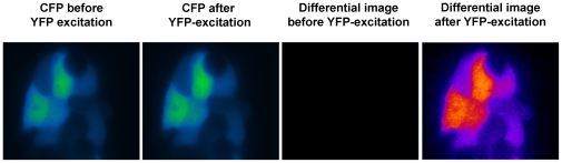
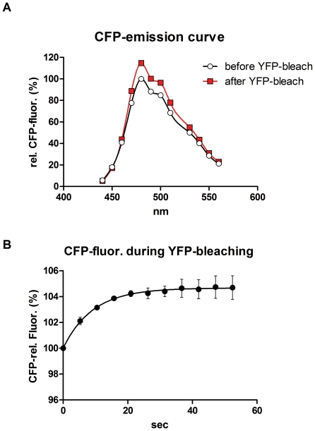
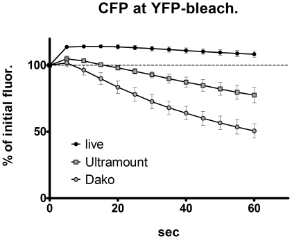
METHODOLOGY/PRINCIPAL FINDINGS: When we applied a commonly used FRET microscopy technique--the increase in donor (CFP)-fluorescence after bleaching of acceptor fluorophores (YFP), we obtained good signals in live cells, but very weak signals for the same samples after fixation and mounting in commercial microscopy mounting fluids. This observation could be traced back to much faster bleaching of CFP in these mounting media. Strikingly, the opposite effect of the mounting fluid was observed for YFP and also for other proteins such as Cerulean, TFP or Venus. The changes in photostability of CFP and YFP were not caused by the fixation but directly dependent on the mounting fluid. Furthermore we made the interesting observation that the CFP-fluorescence intensity increases by about 10-15% after illumination at the YFP-excitation wavelength--a phenomenon, which was also observed for Cerulean. This photoactivation of cyan fluorescent proteins at the YFP-excitation can cause false-positive signals in the FRET-microscopy technique that is based on bleaching of a yellow FRET acceptor.

CONCLUSIONS/SIGNIFICANCE: Our results show that photostability of fluorescent proteins differs significantly for various media and that CFP bleaches significantly faster in commercial mounting fluids, while the opposite is observed for YFP and some other proteins. Moreover, we show that the FRET microscopy technique that is based on bleaching of the YFP is prone to artifacts due to photoactivation of cyan fluorescent proteins under these conditions.

摘要

背景

在生命科学的各个领域,例如标记特定蛋白质或细胞或确定蛋白质相互作用,使用光谱上明显不同的绿色荧光蛋白(GFP)变体(分别为青色或黄色突变体,CFP 和 YFP)非常普遍。在后一种情况下,荧光共振能量转移(FRET)的量子物理现象被特定的显微镜技术利用来可视化蛋白质的接近度。

方法/主要发现:当我们应用一种常用的 FRET 显微镜技术——在猝灭供体荧光团(YFP)的荧光后,CFP 的荧光增加,我们在活细胞中获得了良好的信号,但在固定和安装在商业显微镜安装液中的相同样本中,信号非常弱。这种观察可以追溯到这些安装介质中 CFP 更快的猝灭。引人注目的是,对于 YFP 以及其他蛋白质,如 Cerulean、TFP 或 Venus,安装液具有相反的效果。CFP 和 YFP 光稳定性的变化不是由固定引起的,而是直接取决于安装液。此外,我们还观察到一个有趣的现象,即在 YFP 激发波长下照明后,CFP 的荧光强度增加了约 10-15%——这一现象也在 Cerulean 中观察到。这种在 YFP 激发下的青色荧光蛋白的光激活会导致基于黄色 FRET 受体猝灭的 FRET 显微镜技术中产生假阳性信号。

结论/意义:我们的结果表明,荧光蛋白的光稳定性在各种介质中差异很大,并且 CFP 在商业安装液中猝灭得更快,而 YFP 和其他一些蛋白质则相反。此外,我们还表明,基于 YFP 猝灭的 FRET 显微镜技术由于在这些条件下青色荧光蛋白的光激活,容易产生假象。

https://cdn.ncbi.nlm.nih.gov/pmc/blobs/a20a/3072413/cc2ad8825785/pone.0018586.g008.jpg
https://cdn.ncbi.nlm.nih.gov/pmc/blobs/a20a/3072413/63ebc349b2bc/pone.0018586.g001.jpg
https://cdn.ncbi.nlm.nih.gov/pmc/blobs/a20a/3072413/341d63454588/pone.0018586.g002.jpg
https://cdn.ncbi.nlm.nih.gov/pmc/blobs/a20a/3072413/552620d7cf43/pone.0018586.g003.jpg
https://cdn.ncbi.nlm.nih.gov/pmc/blobs/a20a/3072413/8162c3e8a58b/pone.0018586.g004.jpg
https://cdn.ncbi.nlm.nih.gov/pmc/blobs/a20a/3072413/47baf2b0ba14/pone.0018586.g005.jpg
https://cdn.ncbi.nlm.nih.gov/pmc/blobs/a20a/3072413/a1b3906ac9f0/pone.0018586.g006.jpg
https://cdn.ncbi.nlm.nih.gov/pmc/blobs/a20a/3072413/7af2e13ccf9e/pone.0018586.g007.jpg
https://cdn.ncbi.nlm.nih.gov/pmc/blobs/a20a/3072413/cc2ad8825785/pone.0018586.g008.jpg

相似文献

1
Some secrets of fluorescent proteins: distinct bleaching in various mounting fluids and photoactivation of cyan fluorescent proteins at YFP-excitation.一些荧光蛋白的秘密:在不同的固定液中有明显的漂白现象,以及在 YFP 激发下青色荧光蛋白的光激活。
PLoS One. 2011 Apr 7;6(4):e18586. doi: 10.1371/journal.pone.0018586.
2
Flow cytometric measurement of fluorescence (Förster) resonance energy transfer from cyan fluorescent protein to yellow fluorescent protein using single-laser excitation at 458 nm.利用458nm单激光激发,通过流式细胞术测量从青色荧光蛋白到黄色荧光蛋白的荧光(Förster)共振能量转移。
Cytometry A. 2003 May;53(1):39-54. doi: 10.1002/cyto.a.10037.
3
A flow cytometric method to detect protein-protein interaction in living cells by directly visualizing donor fluorophore quenching during CFP-->YFP fluorescence resonance energy transfer (FRET).一种通过在CFP→YFP荧光共振能量转移(FRET)过程中直接观察供体荧光团淬灭来检测活细胞中蛋白质-蛋白质相互作用的流式细胞术方法。
Cytometry A. 2003 Oct;55(2):71-85. doi: 10.1002/cyto.a.10073.
4
Probing plasma membrane microdomains in cowpea protoplasts using lipidated GFP-fusion proteins and multimode FRET microscopy.利用脂化绿色荧光蛋白融合蛋白和多模式荧光共振能量转移显微镜探测豇豆原生质体中的质膜微区。
J Microsc. 2004 May;214(Pt 2):190-200. doi: 10.1111/j.0022-2720.2004.01318.x.
5
Fluorescence resonance energy transfer from cyan to yellow fluorescent protein detected by acceptor photobleaching using confocal microscopy and a single laser.利用共聚焦显微镜和单激光通过受体光漂白检测从青色荧光蛋白到黄色荧光蛋白的荧光共振能量转移。
J Microsc. 2003 Jan;209(Pt 1):56-70. doi: 10.1046/j.1365-2818.2003.01100.x.
6
Quantifying the influence of yellow fluorescent protein photoconversion on acceptor photobleaching-based fluorescence resonance energy transfer measurements.量化黄色荧光蛋白光转化对基于供体荧光猝灭的荧光共振能量转移测量的影响。
J Biomed Opt. 2012 Jan;17(1):011010. doi: 10.1117/1.JBO.17.1.011010.
7
High-precision FLIM-FRET in fixed and living cells reveals heterogeneity in a simple CFP-YFP fusion protein.在固定细胞和活细胞中进行的高精度荧光寿命成像-荧光共振能量转移(FLIM-FRET)揭示了一种简单的CFP-YFP融合蛋白中的异质性。
Biophys Chem. 2007 May;127(3):155-64. doi: 10.1016/j.bpc.2007.01.008. Epub 2007 Feb 1.
8
Optimization of pairings and detection conditions for measurement of FRET between cyan and yellow fluorescent proteins.用于测量青色和黄色荧光蛋白之间荧光共振能量转移的配对及检测条件的优化。
Microsc Microanal. 2006 Jun;12(3):238-54. doi: 10.1017/S1431927606060235.
9
ATP changes the fluorescence lifetime of cyan fluorescent protein via an interaction with His148.ATP 通过与 His148 的相互作用改变青色荧光蛋白的荧光寿命。
PLoS One. 2010 Nov 5;5(11):e13862. doi: 10.1371/journal.pone.0013862.
10
Resonance energy transfer between green fluorescent protein variants: complexities revealed with myosin fusion proteins.绿色荧光蛋白变体之间的共振能量转移:肌球蛋白融合蛋白揭示的复杂性
Biochemistry. 2006 Sep 5;45(35):10482-91. doi: 10.1021/bi060943u.

引用本文的文献

1
Single-molecule imaging and molecular dynamics simulations reveal early activation of the MET receptor in cells.单分子成像和分子动力学模拟揭示了 MET 受体在细胞中的早期激活。
Nat Commun. 2024 Nov 2;15(1):9486. doi: 10.1038/s41467-024-53772-7.
2
Optimal inference of molecular interaction dynamics in FRET microscopy.在荧光共振能量转移显微镜中对分子相互作用动力学的最佳推断。
Proc Natl Acad Sci U S A. 2023 Apr 11;120(15):e2211807120. doi: 10.1073/pnas.2211807120. Epub 2023 Apr 4.
3
Estimating the Interaction Strength Between PTS1-Peptides and Their Receptor PEX5 in Living Cells Using Flow-Cytometer-Based FRET (flowFRET) Measurements.

本文引用的文献

1
Enhanced yellow fluorescent protein photoconversion to a cyan fluorescent protein-like species is sensitive to thermal and diffusion conditions.增强型黄色荧光蛋白向类青色荧光蛋白物种的光转化对热和扩散条件敏感。
J Biomed Opt. 2009 May-Jun;14(3):034039. doi: 10.1117/1.3103338.
2
Detecting protein-protein interactions with CFP-YFP FRET by acceptor photobleaching.通过受体光漂白利用CFP-YFP荧光共振能量转移检测蛋白质-蛋白质相互作用。
Curr Protoc Cytom. 2006 Feb;Chapter 12:Unit12.7. doi: 10.1002/0471142956.cy1207s35.
3
Fluorescence resonance energy transfer of GFP and YFP by spectral imaging and quantitative acceptor photobleaching.
使用基于流式细胞仪的荧光共振能量转移(flowFRET)测量方法估算 PTS1 肽与其受体 PEX5 在活细胞中的相互作用强度。
Methods Mol Biol. 2023;2643:413-434. doi: 10.1007/978-1-0716-3048-8_30.
4
Antimicrobial Compounds from Microorganisms.微生物来源的抗菌化合物。
Antibiotics (Basel). 2022 Feb 22;11(3):285. doi: 10.3390/antibiotics11030285.
5
Imaging Meets Cytometry: Analyzing Heterogeneous Functional Microscopic Data from Living Cell Populations.成像与细胞计数技术的结合:分析来自活细胞群体的异质性功能显微镜数据。
J Imaging. 2021 Jan 13;7(1):9. doi: 10.3390/jimaging7010009.
6
Flow cytometry-based FRET identifies binding intensities in PPARγ1 protein-protein interactions in living cells.基于流式细胞术的 FRET 可鉴定活细胞中 PPARγ1 蛋白-蛋白相互作用的结合强度。
Theranostics. 2019 Jul 28;9(19):5444-5463. doi: 10.7150/thno.29367. eCollection 2019.
7
Advanced FRET normalization allows quantitative analysis of protein interactions including stoichiometries and relative affinities in living cells.高级 FRET 归一化允许对活细胞中的蛋白质相互作用进行定量分析,包括化学计量和相对亲和力。
Sci Rep. 2019 Jun 3;9(1):8233. doi: 10.1038/s41598-019-44650-0.
8
Improving Quality, Reproducibility, and Usability of FRET-Based Tension Sensors.提高基于 FRET 的张力传感器的质量、可重复性和易用性。
Cytometry A. 2019 Feb;95(2):201-213. doi: 10.1002/cyto.a.23688. Epub 2018 Dec 6.
9
Characterization of MLKL-mediated Plasma Membrane Rupture in Necroptosis.坏死性凋亡中混合谱系激酶结构域样蛋白介导的质膜破裂的特征分析
J Vis Exp. 2018 Aug 7(138):58088. doi: 10.3791/58088.
10
Improving brightness and photostability of green and red fluorescent proteins for live cell imaging and FRET reporting.提高用于活细胞成像和荧光共振能量转移报告的绿色和红色荧光蛋白的亮度及光稳定性。
Sci Rep. 2016 Feb 16;6:20889. doi: 10.1038/srep20889.
通过光谱成像和定量受体光漂白对绿色荧光蛋白(GFP)和黄色荧光蛋白(YFP)进行荧光共振能量转移
J Microsc. 2008 Jul;231(Pt 1):97-104. doi: 10.1111/j.1365-2818.2008.02020.x.
4
Imaging protein-protein interactions by Fluorescence Resonance Energy Transfer (FRET) microscopy.通过荧光共振能量转移(FRET)显微镜成像蛋白质-蛋白质相互作用。
Curr Protoc Neurosci. 2006 Feb;Chapter 5:Unit 5.22. doi: 10.1002/0471142301.ns0522s34.
5
In vivo interactions of MyoD, Id1, and E2A proteins determined by acceptor photobleaching fluorescence resonance energy transfer.通过受体光漂白荧光共振能量转移确定的MyoD、Id1和E2A蛋白的体内相互作用。
FASEB J. 2008 Jun;22(6):1694-701. doi: 10.1096/fj.07-095000. Epub 2008 Jan 15.
6
Sensitive detection of p65 homodimers using red-shifted and fluorescent protein-based FRET couples.使用红移和基于荧光蛋白的荧光共振能量转移对灵敏检测p65同二聚体。
PLoS One. 2007 Oct 10;2(10):e1011. doi: 10.1371/journal.pone.0001011.
7
YFP photoconversion revisited: confirmation of the CFP-like species.再探黄色荧光蛋白光转化:类青色荧光蛋白物种的确认
Nat Methods. 2007 Oct;4(10):767-8. doi: 10.1038/nmeth1007-767.
8
Fluorescent protein FRET: the good, the bad and the ugly.荧光蛋白荧光共振能量转移:优点、缺点与不足
Trends Biochem Sci. 2007 Sep;32(9):407-14. doi: 10.1016/j.tibs.2007.08.003. Epub 2007 Aug 30.
9
Directed evolution of a monomeric, bright and photostable version of Clavularia cyan fluorescent protein: structural characterization and applications in fluorescence imaging.海鸡冠藻青色荧光蛋白单体、明亮且光稳定版本的定向进化:结构表征及其在荧光成像中的应用
Biochem J. 2006 Dec 15;400(3):531-40. doi: 10.1042/BJ20060874.
10
Photobleaching of YFP does not produce a CFP-like species that affects FRET measurements.黄色荧光蛋白(YFP)的光漂白不会产生影响荧光共振能量转移(FRET)测量的类似青色荧光蛋白(CFP)的物种。
Nat Methods. 2006 Jul;3(7):491; author reply 492-3. doi: 10.1038/nmeth0706-491a.