DRK Blood Service Baden-Württemberg-Hessia, Institute for Clinical Transfusion Medicine and Immunogenetics Ulm and Institute of Transfusion Medicine, University of Ulm, Ulm, Germany.
Cytotherapy. 2011 Sep;13(8):962-75. doi: 10.3109/14653249.2011.571246. Epub 2011 Apr 15.
Mesenchymal stromal cells (MSC) are the focus of research in regenerative medicine aiming at the regulatory approval of these cells for specific indications. To cope with the regulatory requirements for somatic cell therapy, novel approaches that do not interfere with the natural behavior of the cells are necessary. In this context in vivo magnetic resonance imaging (MRI) of labeled MSC could be an appropriate tool. Cell labeling for MRI with a variety of different iron oxide preparations is frequently published. However, most publications lack a comprehensive assessment of the non-interference of the contrast agent with the functionality of the labeled MSC, which is a prerequisite for the validity of cell-tracking via MRI.
We studied the effects of iron oxide-poly(l-lactide) nanoparticles in MSC with flow cytometry, transmission electron microscopy (TEM), confocal laser scanning microscopy (CLSM), Prussian blue staining, CyQuant® proliferation testing, colony-forming unit-fibroblast (CFU-F) assays, flow chamber adhesion testing, immunologic tests and differentiation tests. Furthermore iron-labeled MSC were studied by MRI in agarose phantoms and Wistar rats.
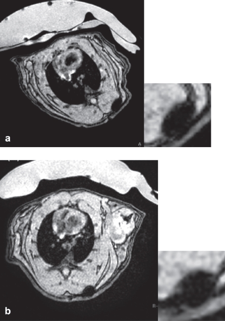
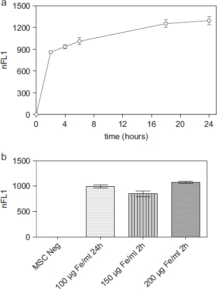
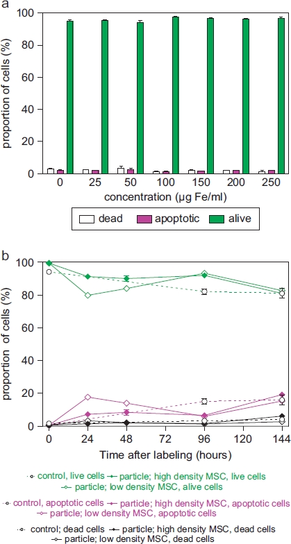
It could be demonstrated that MSC show rapid uptake of nanoparticles and long-lasting intracellular persistence in the endosomal compartment. Labeling of the MSC with these particles has no influence on viability, differentiation, clonogenicity, proliferation, adhesion, phenotype and immunosuppressive properties. They show excellent MRI properties in agarose phantoms and after subcutaneous implantation in rats over several weeks.
These particles qualify for studying MSC homing and trafficking via MRI.
间充质基质细胞(MSC)是再生医学研究的焦点,旨在使这些细胞获得特定适应症的监管批准。为了满足体细胞治疗的监管要求,有必要采用不会干扰细胞自然行为的新方法。在这种情况下,体内磁共振成像(MRI)对标记的 MSC 进行成像可能是一种合适的工具。用各种不同的氧化铁制剂对细胞进行 MRI 标记经常被报道。然而,大多数出版物缺乏对造影剂对标记的 MSC 功能的非干扰的全面评估,这是通过 MRI 进行细胞追踪的有效性的前提。
我们使用流式细胞术、透射电子显微镜(TEM)、共聚焦激光扫描显微镜(CLSM)、普鲁士蓝染色、CyQuant®增殖检测、集落形成单位-成纤维细胞(CFU-F)检测、流动室粘附试验、免疫试验和分化试验研究了氧化铁-聚(L-丙交酯)纳米粒子对 MSC 的影响。此外,还在琼脂糖体模和 Wistar 大鼠中研究了铁标记的 MSC 的 MRI。
可以证明 MSC 迅速摄取纳米颗粒,并在内涵体隔室中持久存在。用这些颗粒对 MSC 进行标记不会影响其活力、分化、集落形成能力、增殖、粘附、表型和免疫抑制特性。它们在琼脂糖体模和大鼠皮下植入数周后表现出优异的 MRI 特性。
这些颗粒有资格通过 MRI 研究 MSC 归巢和迁移。