Department of Radiology, Medical University of Gdansk, Debinki 7, 80-211 Gdansk, Poland.
BMC Gastroenterol. 2011 Apr 19;11:43. doi: 10.1186/1471-230X-11-43.
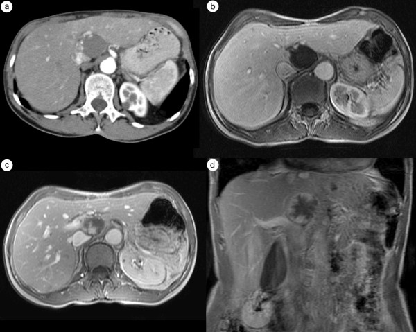
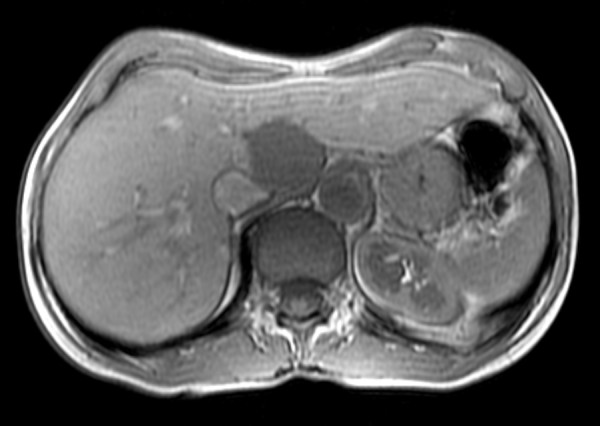
Cavernous hemangiomas are the most frequent neoplasms of the liver and in routine clinical practice they often need to be differentiated from malignant tumors and other benign focal lesions. The purpose of this study is to evaluate whether diagnostic accuracy of magnetic resonance imaging (MRI) of hepatic hemangiomas, showing atypical pattern on US, improves with the use of Gd-BOPTA in comparison with contrast-enhanced multi-phase computed tomography (CT).
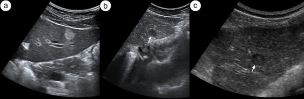
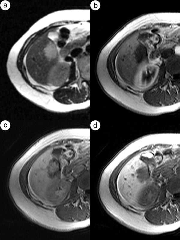
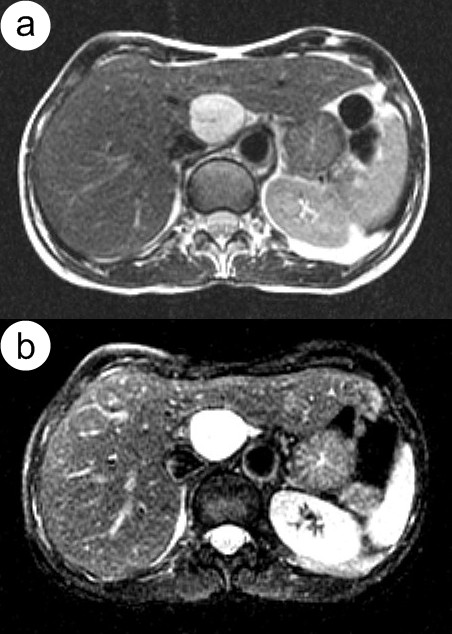
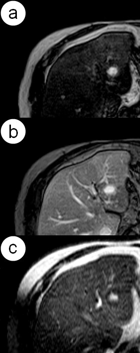
178 consecutive patients with ambiguous hepatic masses showing atypical hyperechoic pattern on grey-scale US, underwent unenhanced and contrast-enhanced multi-phase multi-detector CT and MR (1.5T) with the use of liver-specific contrast medium gadobenate dimeglumine (Gd-BOPTA). After intravenous contrast administration arterial (HAP), venous-portal (PVP), equilibrium phases (EP) both in CT and MR and additionally hepatobiliary phase (HBP) in MR were obtained. 398 lesions have been detected including 99 hemangiomas and 299 other lesions.
In non-enhanced MDCT examination detection of hemangiomas was characterized by sensitivity of 76%, specificity of 90%, PPV of 71%, NPV of 92% and accuracy of 86%.Non-enhanced MR examination showed sensitivity of 98%, specificity of 99%, PPV of 99%, NPV of 99% and accuracy of 99%.After intravenous administration of contrast medium in MR the mentioned above parameters did not increase significantly.
Gd-BOPTA-enhanced MR in comparison with unenhanced MRI does not improve diagnostic accuracy in discriminating hemangiomas that show non-specific appearance in ultrasound examination. Unenhanced MR as a method of choice should directly follow US in course of diagnostic algorithm in differentiation of hemangiomas from other liver tumors.
海绵状血管瘤是肝脏最常见的肿瘤,在常规临床实践中,它们经常需要与恶性肿瘤和其他良性局灶性病变区分开来。本研究的目的是评估在超声显示非典型回声模式的情况下,磁共振成像(MRI)对肝血管瘤的诊断准确性是否通过使用钆塞酸二钠(Gd-BOPTA)与多期增强 CT(CT)相比有所提高。
178 例连续患者的肝脏肿块在灰阶超声上显示非典型高回声模式,接受了未增强和多期多探测器 CT 和 MRI(1.5T)检查,使用了肝特异性造影剂钆塞酸二钠(Gd-BOPTA)。静脉注射对比剂后,在 CT 和 MRI 上分别获得了动脉期(HAP)、门静脉期(PVP)、平衡期(EP),以及 MRI 上的肝胆期(HBP)。共检测到 398 个病灶,包括 99 个血管瘤和 299 个其他病灶。
在非增强 MDCT 检查中,检测到的血管瘤的敏感性为 76%,特异性为 90%,PPV 为 71%,NPV 为 92%,准确性为 86%。非增强 MRI 检查显示敏感性为 98%,特异性为 99%,PPV 为 99%,NPV 为 99%,准确性为 99%。在 MRI 中静脉注射对比剂后,上述参数没有显著增加。
与未增强 MRI 相比,钆塞酸二钠增强 MRI 并不能提高在超声检查中显示非特异性表现的血管瘤的诊断准确性。作为首选方法的未增强 MRI 应在超声检查后直接用于诊断算法,以区分血管瘤与其他肝脏肿瘤。