Suppr超能文献

外周巨细胞肉芽肿:123例病例回顾

Peripheral giant cell granuloma: a review of 123 cases.

作者信息

Shadman Niloofar, Ebrahimi Shahram Farzin, Jafari Shahin, Eslami Mohammad

机构信息

Assistant professor, Department of Restorative Dentistry, School of Dentistry, Kerman University of Medical Sciences, Kerman, Iran.

出版信息

Dent Res J (Isfahan). 2009 Spring;6(1):47-50.

Abstract

BACKGROUND

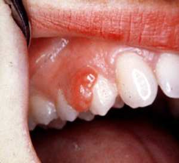
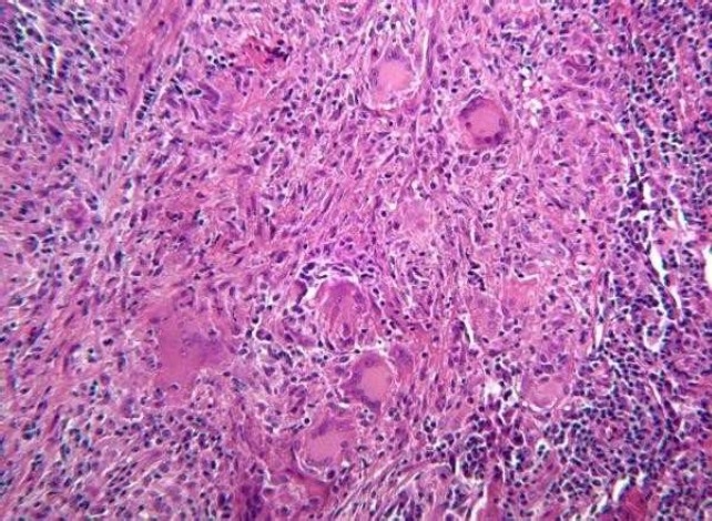
Peripheral giant cell granuloma is one of the reactive hyperplastic lesions of the oral cavity, which originates from the periosteum or periodontal membrane following local irritation or chronic trauma. The purpose of this study was to present the clinical characteristics of peripheral giant cell granuloma in a group of Iranian population.

METHODS

A series of 123 consecutive confirmed cases of peripheral giant cell granuloma after biopsy were evaluated. Age, sex, anatomic location, consistency, etiologic factor, pain and bleeding history, color, surface texture, and pedicle situation were recorded and were analyzed by chi-square test and values were considered to be significant if P < 0.05.

RESULTS

Age ranged from 6 to 75 years (mean 33 years). Women affected more than men (M/F 1:1.1). Peripheral giant cell granuloma was seen in the mandible more than in the maxilla and in the anterior region more than in the posterior region. In most cases, lesions were pink, pedunculated and had non-ulcerated surface. In less than half of the cases, there was no history of bleeding and also pain was rarely reported. Calculus was the most common etiologic factor.

CONCLUSION

The results confirmed that the clinical features of peripheral giant cell granuloma in a group of Iranian population are almost similar to those reported by other investigators.

摘要

背景

外周性巨细胞肉芽肿是口腔的反应性增生性病变之一,其起源于局部刺激或慢性创伤后的骨膜或牙周膜。本研究的目的是呈现一组伊朗人群中外周性巨细胞肉芽肿的临床特征。

方法

对一系列123例经活检确诊的外周性巨细胞肉芽肿连续病例进行评估。记录年龄、性别、解剖位置、质地、病因、疼痛和出血史、颜色、表面质地和蒂部情况,并通过卡方检验进行分析,若P < 0.05,则认为差异具有统计学意义。

结果

年龄范围为6至75岁(平均33岁)。女性受累多于男性(男/女为1:1.1)。外周性巨细胞肉芽肿在下颌骨比在上颌骨更常见,在前部区域比在后部区域更常见。在大多数情况下,病变为粉红色、有蒂且表面无溃疡。不到一半的病例有出血史,疼痛也很少被报告。牙结石是最常见的病因。

结论

结果证实,一组伊朗人群中外周性巨细胞肉芽肿的临床特征与其他研究者报告的特征几乎相似。

https://cdn.ncbi.nlm.nih.gov/pmc/blobs/ae98/3075451/f0d05aa9aa0a/DRJ-6-47-g001.jpg

相似文献

1
Peripheral giant cell granuloma: a review of 123 cases.
Dent Res J (Isfahan). 2009 Spring;6(1):47-50.
2
Reactive hyperplasia of the oral cavity in Kerman province, Iran: a review of 172 cases.
Br J Oral Maxillofac Surg. 2007 Jun;45(4):288-92. doi: 10.1016/j.bjoms.2006.10.001. Epub 2006 Nov 9.
3
Surgical excision of peripheral giant cell granuloma of the maxilla: a case report.
Pan Afr Med J. 2023 Mar 20;44:141. doi: 10.11604/pamj.2023.44.141.34835. eCollection 2023.
4
An Audit of Histopathological Pattern of Peripheral Giant Cell Granuloma - A Retrospective Study.
Ann Maxillofac Surg. 2024 Jan-Jun;14(1):62-65. doi: 10.4103/ams.ams_20_24. Epub 2024 Jul 19.
5
Giant cell epulis.
Wien Med Wochenschr. 2023 Sep;173(11-12):249-250. doi: 10.1007/s10354-021-00894-y. Epub 2021 Nov 4.
6
Peripheral giant cell granuloma.
Contemp Clin Dent. 2012 Apr;3(Suppl 1):S118-21. doi: 10.4103/0976-237X.95121.
7
A Review and Report of Peripheral Giant Cell Granuloma in a 4-Year-Old Child.
Case Rep Dent. 2016;2016:7536304. doi: 10.1155/2016/7536304. Epub 2016 Jun 15.
9
Peripheral giant cell granuloma associated with dental implants.
J Periodontol. 2003 Sep;74(9):1381-4. doi: 10.1902/jop.2003.74.9.1381.
10
Management of an unusual peripheral giant cell granuloma: A diagnostic dilemma.
Contemp Clin Dent. 2012 Jan;3(1):93-6. doi: 10.4103/0976-237X.94556.

引用本文的文献

1
Expansive Oral Giant Cell Granuloma in a Pediatric Patient.
Int J Clin Pediatr Dent. 2023 Mar-Apr;16(2):405-408. doi: 10.5005/jp-journals-10005-2572.
2
Peripheral giant cell granuloma: a case series and brief review.
Ann R Coll Surg Engl. 2024 Sep;106(7):649-651. doi: 10.1308/rcsann.2023.0021. Epub 2023 Jun 27.
4
Peripheral giant cell granuloma of maxilla.
J Indian Soc Periodontol. 2022 Jan-Feb;26(1):75-78. doi: 10.4103/jisp.jisp_624_20. Epub 2021 Sep 27.
6
A Nervous Recipient of a "Tongue Lashing".
Fed Pract. 2020 Mar;37(3):146-148.
8
Peripheral and Central Giant Cell Lesions in Children: Institutional Experience at Subharti Dental College and Hospital.
Indian J Med Paediatr Oncol. 2017 Oct-Dec;38(4):440-446. doi: 10.4103/ijmpo.ijmpo_17_16.
10
A Review and Report of Peripheral Giant Cell Granuloma in a 4-Year-Old Child.
Case Rep Dent. 2016;2016:7536304. doi: 10.1155/2016/7536304. Epub 2016 Jun 15.

本文引用的文献

2
Peripheral and central giant cell granulomas of the jaws: a demographic study.
Oral Surg Oral Med Oral Pathol Oral Radiol Endod. 2007 Jun;103(6):e39-43. doi: 10.1016/j.tripleo.2006.12.022. Epub 2007 Apr 11.
4
Peripheral giant cell granuloma. A report of five cases and review of the literature.
Med Oral Patol Oral Cir Bucal. 2005 Jan-Feb;10(1):53-7; 48-52.
5
8
The peripheral giant cell granuloma.
Gen Dent. 1997 Nov-Dec;45(6):577-9.
9
Growth potential of peripheral giant cell granuloma.
Oral Surg Oral Med Oral Pathol Oral Radiol Endod. 1997 May;83(5):548-51. doi: 10.1016/s1079-2104(97)90118-9.
10
Peripheral giant-cell granuloma: a review.
J Ir Dent Assoc. 1994;40(2):39-41.

文献AI研究员

20分钟写一篇综述,助力文献阅读效率提升50倍。

立即体验

用中文搜PubMed

大模型驱动的PubMed中文搜索引擎

马上搜索

文档翻译

学术文献翻译模型,支持多种主流文档格式。

立即体验