Laboratory of Pathology, Instituto Oswaldo Cruz/Fiocruz, Av. Brasil 4365, Pavilhão Gomes de Faria, Rio de Janeiro, CEP 21040-360, Brazil.
Cell Tissue Res. 2011 Jun;344(3):455-69. doi: 10.1007/s00441-011-1170-1. Epub 2011 May 4.
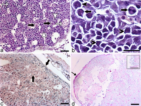
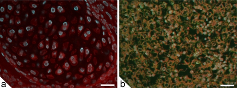
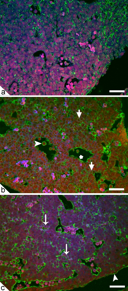
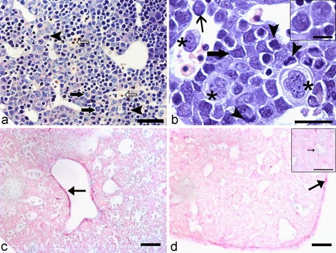
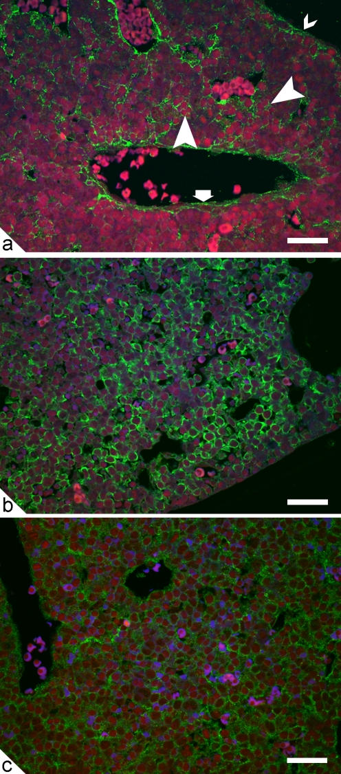
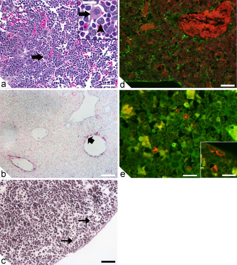
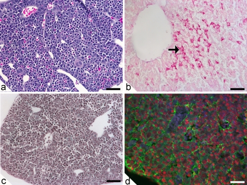
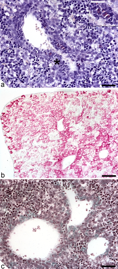
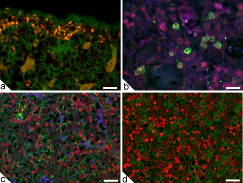
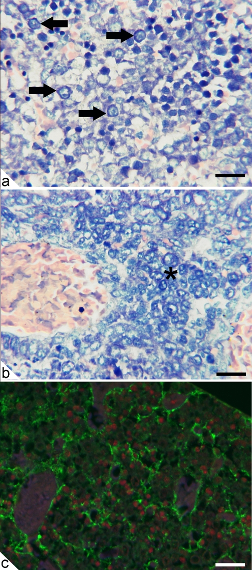
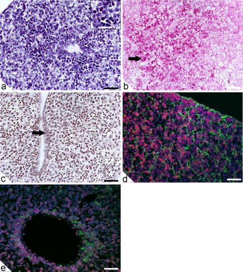
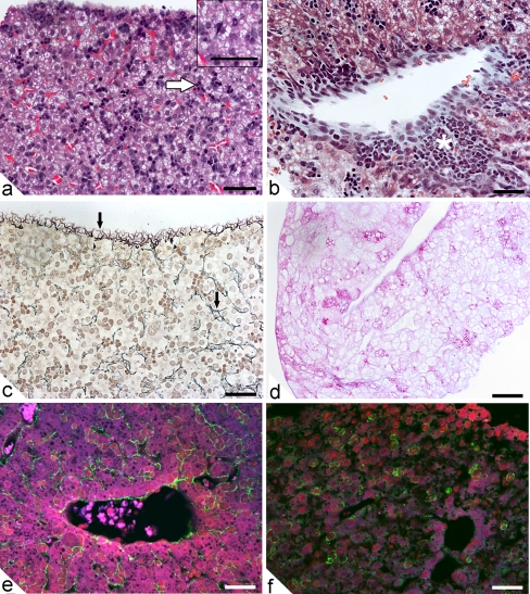
Embryonic hematopoiesis occurs via dynamic development with cells migrating into various organs. Fetal liver is the main hematopoietic organ responsible for hematopoietic cell expansion during embryologic development. We describe the morphological sequential characteristics of murine fetal liver niches that favor the settlement and migration of hematopoietic cells from 12 days post-coitum (dpc) to 0 day post-partum. Liver sections were stained with hematoxylin and eosin, Lennert's Giemsa, Sirius Red pH 10.2, Gomori's Reticulin, and Periodic Acid Schiff/Alcian Blue pH 1.0 and pH 2.5 and were analyzed by bright-field microscopy. Indirect imunohistochemistry for fibronectin, matrix metalloproteinase-1 (MMP-1), and MMP-9 and histochemistry for naphthol AS-D chloroacetate esterase (NCAE) were analyzed by confocal microscopy. The results showed that fibronectin was related to the promotion of hepatocyte and trabecular differentiation; reticular fibers did not appear to participate in fetal hematopoiesis but contributed to the physical support of the liver after 18 dpc. During the immature phase, hepatocytes acted as the fundamental stroma for the erythroid lineage. The appearance of myeloid cells in the liver was related to perivascular and subcapsular collagen, and NCAE preceded MMP-1 expression in neutrophils, an occurrence that appeared to contribute to their liver evasion. Thus, the murine fetal liver during ontogenesis shows two different phases: one immature and mainly endodermic (<14 dpc) and the other more developed (endodermic-mesenchymal; >15 dpc) with the maturation of hepatocytes, a better definition of trabecular pattern, and an increase in the connective tissue in the capsule, portal spaces, and liver parenchyma. The decrease of hepatic hematopoiesis (migration) coincides with hepatic maturation.
胚胎造血发生通过细胞迁移到各种器官的动态发育进行。胎儿肝脏是胚胎发育过程中负责造血细胞扩增的主要造血器官。我们描述了有利于造血细胞从 12 天妊娠龄(post-coitum,dpc)到 0 天产后定居和迁移的鼠胎儿肝脏龛的形态顺序特征。肝切片用苏木精和伊红、Lennert 的 Giemsa、Sirius Red pH 10.2、Gomori 的网状纤维、过碘酸雪夫/阿尔辛蓝 pH 1.0 和 pH 2.5 染色,并通过明场显微镜进行分析。纤维连接蛋白、基质金属蛋白酶-1(MMP-1)和 MMP-9 的间接免疫组织化学和萘酚 AS-D 氯乙酸酯酶(NCAE)的组织化学通过共聚焦显微镜进行分析。结果表明,纤维连接蛋白与促进肝细胞和小梁分化有关;网状纤维似乎不参与胎儿造血,但在 18 天妊娠龄后有助于肝脏的物理支撑。在不成熟阶段,肝细胞作为红系谱系的基本基质。肝脏中髓样细胞的出现与血管周围和包膜下胶原有关,NCAE 先于中性粒细胞中 MMP-1 的表达,这似乎有助于它们逃避肝脏。因此,在发生过程中,鼠胎儿肝脏表现出两个不同的阶段:一个不成熟且主要是内胚层(<14 dpc),另一个更发达(内胚层-中胚层;>15 dpc),伴随着肝细胞的成熟,小梁模式的更好定义,以及包膜、门腔和肝实质中结缔组织的增加。肝造血(迁移)的减少与肝成熟相吻合。