• 文献检索
  • 文档翻译
  • 深度研究
  • 学术资讯
  • Suppr Zotero 插件Zotero 插件
  • 邀请有礼
  • 套餐&价格
  • 历史记录
应用&插件
Suppr Zotero 插件Zotero 插件浏览器插件Mac 客户端Windows 客户端微信小程序
定价
高级版会员购买积分包购买API积分包
服务
文献检索文档翻译深度研究API 文档MCP 服务
关于我们
关于 Suppr公司介绍联系我们用户协议隐私条款
关注我们

Suppr 超能文献

核心技术专利:CN118964589B侵权必究
粤ICP备2023148730 号-1Suppr @ 2026

文献检索

告别复杂PubMed语法,用中文像聊天一样搜索,搜遍4000万医学文献。AI智能推荐,让科研检索更轻松。

立即免费搜索

文件翻译

保留排版,准确专业,支持PDF/Word/PPT等文件格式,支持 12+语言互译。

免费翻译文档

深度研究

AI帮你快速写综述,25分钟生成高质量综述,智能提取关键信息,辅助科研写作。

立即免费体验

实验鼠的肝外髓外造血。

The hepatic extramedullary hematopoiesis during experimental murine .

机构信息

Laboratory of Pathology, Instituto Oswaldo Cruz, Fiocruz, Rio de Janeiro, Brazil.

Brazilian National Institute of Science and Technology on Neuroimmunomodulation, Oswaldo Cruz Institute, Oswaldo Cruz Foundation, Rio de Janeiro, Brazil.

出版信息

Front Immunol. 2022 Aug 25;13:955034. doi: 10.3389/fimmu.2022.955034. eCollection 2022.

DOI:10.3389/fimmu.2022.955034
PMID:36091027
原文链接:https://pmc.ncbi.nlm.nih.gov/articles/PMC9453041/
Abstract

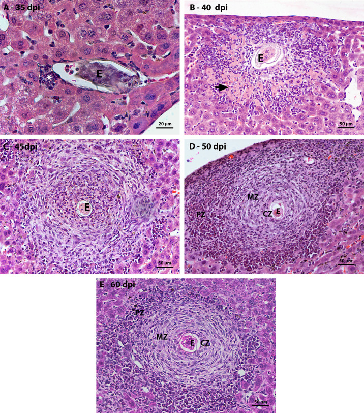
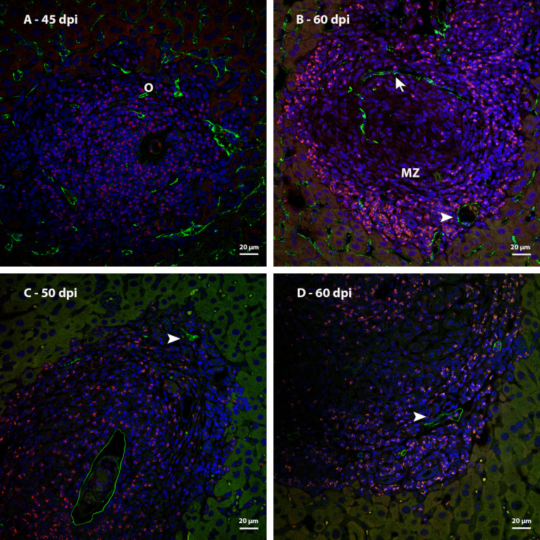
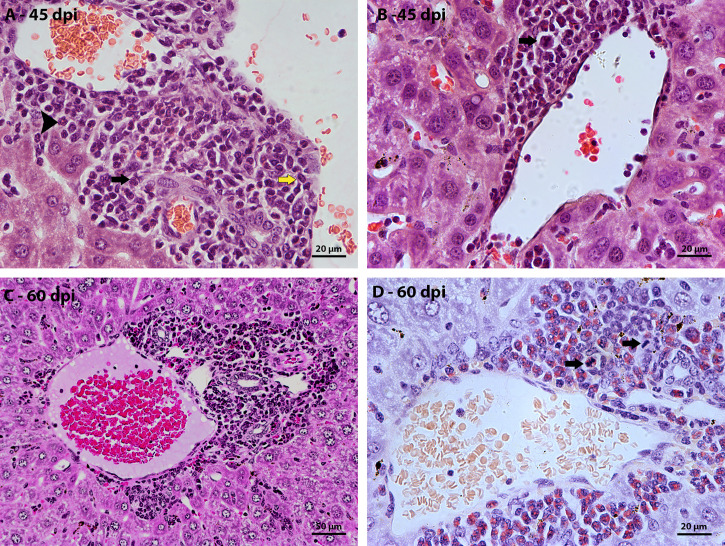
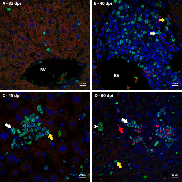
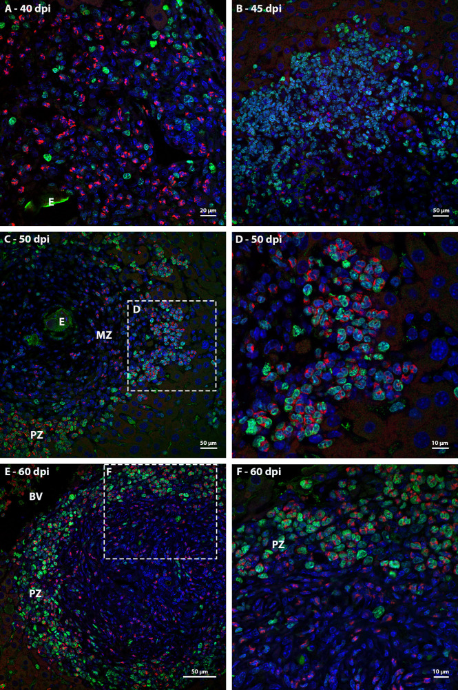
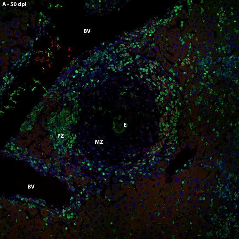
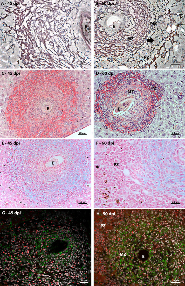
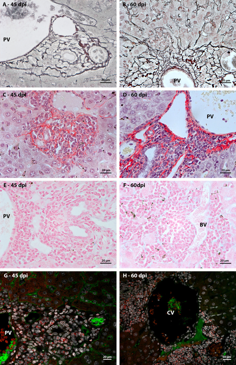
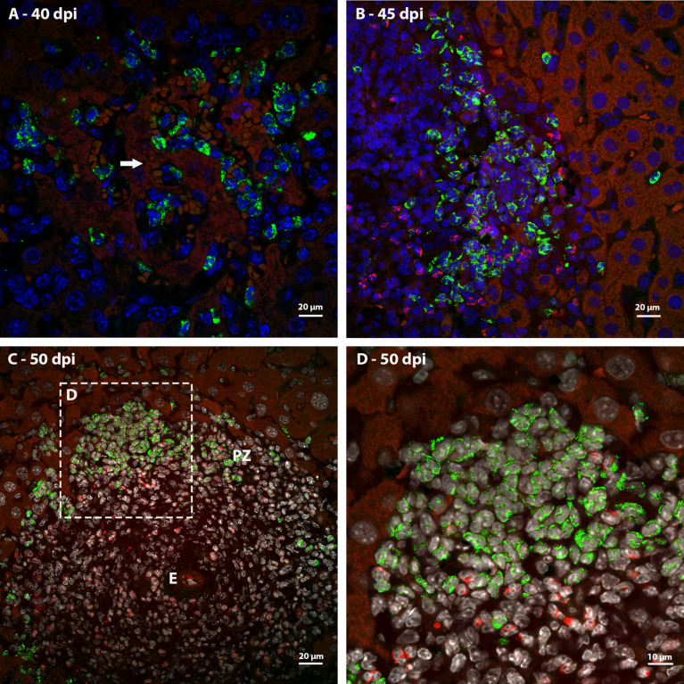
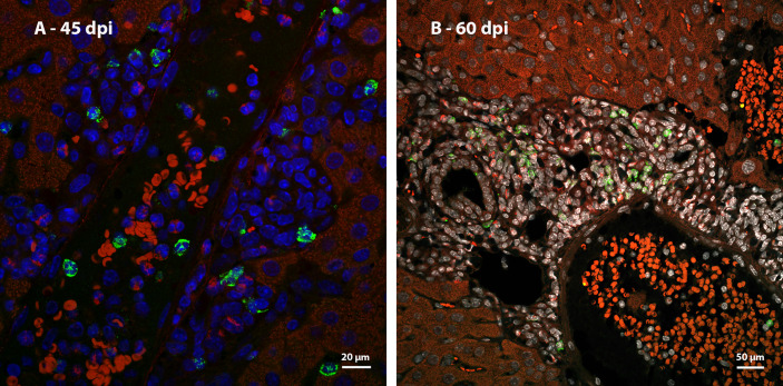
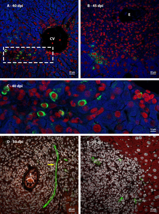
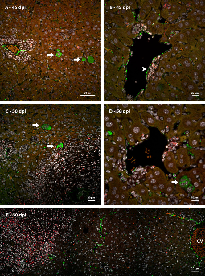
Many years ago, our research group has demonstrated extramedullary hematopoiesis in the peripheral zone of murine hepatic schistosomal granulomas. In the present study, we revisit this phenomenon using new technical and conceptual approaches. Therefore, newborn mice were percutaneously infected by cercariae and euthanized between 35- and 60-days post infection. Liver samples were submitted to histopathology and immunohistochemical analyses. Cells under mitosis and/or expressing Ki67 demonstrated the proliferation of hematopoietic cells both around the parasite's eggs trapped in the liver and around hepatic vessels. After 50 days post infection, proliferating cells at different levels on differentiation were located preferentially in the peripheral zone of the granulomas, around the vessels and inside the sinusoids. The presence of acidic and sulfated glycoconjugates, reticular fibers and the absence of fibronectin characterized the microenvironment for attraction and maintenance of hematopoiesis. Some neutrophils secreted MMP9 from the earliest points of infection, indicating degradation of the extracellular matrix in regions of histolysis and a possible chemoattraction of hematopoietic stem cells to the liver. Fall-3+ cells and Sca-1+ cells indicated that early hematopoietic progenitors could be mobilized to the liver. Groups of vWF+ megakaryocytes suggest chemoattraction of these cells and/or migration, proliferation, and differentiation of very immature progenitors to this organ. The increase of blood vessels and extramedullary hematopoiesis in this environment, where markers of immature hematopoietic and endothelial cells have been identified, points to the possibility of the presence of progenitors for endothelial and hematopoietic cells in the liver during the infection. There is also the possibility of concomitant migration of more differentiated hematopoietic progenitors, that proliferate and differentiate in the liver, and the occurrence of angiogenesis caused by inflammation or release of ovular antigens that stimulate the activation and proliferation of endothelial cells. Altogether, these data increase knowledge about a murine model that is of interest for investigating the pathology of the schistosomiasis and also the dynamics of hematopoiesis.

摘要

许多年前,我们的研究小组已经证明了在鼠类肝血吸虫肉芽肿的外周区存在骨髓外造血。在本研究中,我们使用新的技术和概念方法重新研究了这一现象。因此,新生小鼠经皮感染尾蚴,并在感染后 35-60 天内安乐死。肝组织标本进行组织病理学和免疫组织化学分析。处于有丝分裂和/或表达 Ki67 的细胞表明,在寄生虫卵被困在肝脏和肝血管周围的造血细胞增殖。感染后 50 天,处于不同分化水平的增殖细胞优先位于肉芽肿的外周区,围绕血管和窦内。酸性和硫酸化糖缀合物、网状纤维的存在以及纤连蛋白的缺乏,构成了吸引和维持造血的微环境特征。一些中性粒细胞从感染的最早点开始分泌 MMP9,表明在组织溶解区域的细胞外基质降解,以及造血干细胞向肝脏的可能趋化作用。Fall-3+细胞和 Sca-1+细胞表明,早期造血祖细胞可能被动员到肝脏。vWF+巨核细胞的聚集提示这些细胞的趋化作用,以及/或非常不成熟祖细胞向该器官的迁移、增殖和分化。在这个环境中,血管和骨髓外造血增加,其中已经鉴定出不成熟造血和内皮细胞的标志物,这表明在感染期间,肝脏中可能存在内皮细胞和造血细胞的祖细胞。也有可能同时迁移更分化的造血祖细胞,这些细胞在肝脏中增殖和分化,以及由炎症或卵抗原释放引起的血管生成,刺激内皮细胞的激活和增殖。总之,这些数据增加了对一种感兴趣的鼠类模型的认识,该模型可用于研究血吸虫病的病理学以及造血动力学。

https://cdn.ncbi.nlm.nih.gov/pmc/blobs/4506/9453041/91a600b28759/fimmu-13-955034-g012.jpg
https://cdn.ncbi.nlm.nih.gov/pmc/blobs/4506/9453041/c99f12be6c84/fimmu-13-955034-g001.jpg
https://cdn.ncbi.nlm.nih.gov/pmc/blobs/4506/9453041/d7500110be71/fimmu-13-955034-g002.jpg
https://cdn.ncbi.nlm.nih.gov/pmc/blobs/4506/9453041/fb2461caf1bb/fimmu-13-955034-g003.jpg
https://cdn.ncbi.nlm.nih.gov/pmc/blobs/4506/9453041/e81857fbb911/fimmu-13-955034-g004.jpg
https://cdn.ncbi.nlm.nih.gov/pmc/blobs/4506/9453041/704a10f9d6dd/fimmu-13-955034-g005.jpg
https://cdn.ncbi.nlm.nih.gov/pmc/blobs/4506/9453041/f11c78978081/fimmu-13-955034-g006.jpg
https://cdn.ncbi.nlm.nih.gov/pmc/blobs/4506/9453041/7432b8b9bf62/fimmu-13-955034-g007.jpg
https://cdn.ncbi.nlm.nih.gov/pmc/blobs/4506/9453041/7b0070b0ed5a/fimmu-13-955034-g008.jpg
https://cdn.ncbi.nlm.nih.gov/pmc/blobs/4506/9453041/2c59f57ae61c/fimmu-13-955034-g009.jpg
https://cdn.ncbi.nlm.nih.gov/pmc/blobs/4506/9453041/fbc6d4dc2b62/fimmu-13-955034-g010.jpg
https://cdn.ncbi.nlm.nih.gov/pmc/blobs/4506/9453041/ca457cf14872/fimmu-13-955034-g011.jpg
https://cdn.ncbi.nlm.nih.gov/pmc/blobs/4506/9453041/91a600b28759/fimmu-13-955034-g012.jpg

相似文献

1
The hepatic extramedullary hematopoiesis during experimental murine .实验鼠的肝外髓外造血。
Front Immunol. 2022 Aug 25;13:955034. doi: 10.3389/fimmu.2022.955034. eCollection 2022.
2
Extramedullary hematopoiesis in murine schistosomiasis mansoni.
Mem Inst Oswaldo Cruz. 1995 Mar-Apr;90(2):169-77. doi: 10.1590/s0074-02761995000200008.
3
The host's genetic background determines the extent of angiogenesis induced by schistosome egg antigens.宿主的遗传背景决定了血吸虫卵抗原诱导的血管生成程度。
Acta Trop. 2006 Oct;99(2-3):243-51. doi: 10.1016/j.actatropica.2006.08.011. Epub 2006 Sep 27.
4
Oral Acute Infection in Mice Targets Primary Lymphoid Organs and Triggers Extramedullary Hematopoiesis.口腔急性感染以小鼠的初级淋巴器官为靶标,并引发骨髓外造血。
Front Cell Infect Microbiol. 2022 Mar 24;12:800395. doi: 10.3389/fcimb.2022.800395. eCollection 2022.
5
Hepatic extramedullary hematopoiesis and macrophages in the adult mouse: histometrical and immunohistochemical studies.成年小鼠肝脏骨髓外造血与巨噬细胞:组织计量学和免疫组织化学研究。
Cells Tissues Organs. 2012;196(6):555-64. doi: 10.1159/000338336. Epub 2012 Jun 27.
6
Extramedullary hematopoiesis in the adult mouse liver is associated with specific hepatic sinusoidal endothelial cells.成年小鼠肝脏中的髓外造血与特定的肝窦内皮细胞有关。
Hepatology. 1997 Jul;26(1):165-75. doi: 10.1002/hep.510260122.
7
Extramedullary eosinophilopoiesis in the liver of Schistosoma japonicum-infected mice, with reference to hemopoietic stem cells.
Parasitol Res. 1990;76(6):461-5. doi: 10.1007/BF00931050.
8
Impact of treatment with a Protein Tyrosine Kinase Inhibitor (Genistein) on acute and chronic experimental Schistosoma mansoni infection.蛋白酪氨酸激酶抑制剂(金雀异黄素)治疗对急性和慢性曼氏血吸虫实验性感染的影响。
Exp Parasitol. 2018 Feb;185:115-123. doi: 10.1016/j.exppara.2018.01.013. Epub 2018 Jan 10.
9
The IL-33/ST2 pathway is not essential to Th2 stimulation but is key for modulation and survival during chronic infection with Schistosoma mansoni in mice.IL-33/ST2 通路对于 Th2 刺激并非必需,但对于在感染曼氏血吸虫的慢性感染过程中的调节和存活是关键的。
Cytokine. 2021 Feb;138:155390. doi: 10.1016/j.cyto.2020.155390. Epub 2020 Dec 16.
10
CD18 Regulates Monocyte Hematopoiesis and Promotes Resistance to Experimental Schistosomiasis.CD18 调控单核细胞造血并促进实验性血吸虫病的抵抗。
Front Immunol. 2018 Aug 31;9:1970. doi: 10.3389/fimmu.2018.01970. eCollection 2018.

引用本文的文献

1
A major ecological niche of eosinophils in evolving granulomas challenges the eosinophil view as "helminth killer" cells.嗜酸性粒细胞在不断演变的肉芽肿中的一个主要生态位挑战了将嗜酸性粒细胞视为“杀蠕虫细胞”的观点。
Sci Adv. 2025 Jun 13;11(24):eadt2779. doi: 10.1126/sciadv.adt2779. Epub 2025 Jun 11.
2
Proliferation of MDSCs may indicate a lower  T cell immune response in schistosomiasis japonica.骨髓源性抑制细胞的增殖可能表明日本血吸虫病中的 T 细胞免疫应答较低。
Parasite. 2024;31:52. doi: 10.1051/parasite/2024050. Epub 2024 Aug 29.
3
Schistosome Infection Impacts Hematopoiesis.

本文引用的文献

1
Changing our view of the Schistosoma granuloma to an ecological standpoint.将血吸虫肉芽肿的观点转变为生态立场。
Biol Rev Camb Philos Soc. 2021 Aug;96(4):1404-1420. doi: 10.1111/brv.12708. Epub 2021 Mar 22.
2
Fibronectin and Its Receptors in Hematopoiesis.纤维连接蛋白及其在造血中的受体。
Cells. 2020 Dec 18;9(12):2717. doi: 10.3390/cells9122717.
3
Matrix metalloproteinase-9 (MMP-9) and its inhibitors in cancer: A minireview.基质金属蛋白酶-9(MMP-9)及其在癌症中的抑制剂:综述
血吸虫感染影响造血。
J Immunol. 2024 Feb 15;212(4):607-616. doi: 10.4049/jimmunol.2300195.
4
Modulation of haematopoiesis by protozoal and helminth parasites.原虫和蠕虫寄生虫对造血的调节。
Parasite Immunol. 2023 Dec;45(12):e12975. doi: 10.1111/pim.12975. Epub 2023 Feb 28.
5
Schistosomiasis Mansoni-Recruited Eosinophils: An Overview in the Granuloma Context.曼氏血吸虫病招募的嗜酸性粒细胞:肉芽肿背景概述
Microorganisms. 2022 Oct 13;10(10):2022. doi: 10.3390/microorganisms10102022.
Eur J Med Chem. 2020 May 15;194:112260. doi: 10.1016/j.ejmech.2020.112260. Epub 2020 Mar 21.
4
The mechanisms of pathological extramedullary hematopoiesis in diseases.疾病中病理性骨髓外造血的机制。
Cell Mol Life Sci. 2020 Jul;77(14):2723-2738. doi: 10.1007/s00018-020-03450-w. Epub 2020 Jan 23.
5
New paradigms on hematopoietic stem cell differentiation.造血干细胞分化的新范式。
Protein Cell. 2020 Jan;11(1):34-44. doi: 10.1007/s13238-019-0633-0. Epub 2019 Jun 14.
6
von Willebrand factor regulation of blood vessel formation.血管生成中 von Willebrand 因子的调节。
Blood. 2018 Jul 12;132(2):132-140. doi: 10.1182/blood-2018-01-769018. Epub 2018 Jun 4.
7
Hematopoietic stem cells can differentiate into restricted myeloid progenitors before cell division in mice.造血干细胞在细胞分裂前可在小鼠中分化为受限的髓系祖细胞。
Nat Commun. 2018 May 15;9(1):1898. doi: 10.1038/s41467-018-04188-7.
8
Histological assessment of granulomas in natural and experimental Schistosoma mansoni infections using whole slide imaging.使用全玻片成像技术对自然感染和实验性曼氏血吸虫感染中的肉芽肿进行组织学评估。
PLoS One. 2017 Sep 13;12(9):e0184696. doi: 10.1371/journal.pone.0184696. eCollection 2017.
9
Heparan sulfate proteoglycans as key regulators of the mesenchymal niche of hematopoietic stem cells.硫酸乙酰肝素蛋白聚糖作为造血干细胞间充质微环境的关键调节因子。
Glycoconj J. 2017 Jun;34(3):377-391. doi: 10.1007/s10719-017-9773-8. Epub 2017 Jun 2.
10
Liver inflammation and fibrosis.肝脏炎症和纤维化。
J Clin Invest. 2017 Jan 3;127(1):55-64. doi: 10.1172/JCI88881.