Bellner Lars, Patil Kiran A, Castellano Kirkland, Halilovic Adna, Dunn Michael W, Schwartzman Michal Laniado
Department of Pharmacology, New York Medical College, Valhalla, NY 10595, USA.
Mol Vis. 2011 Apr 29;17:1144-52.
Heme oxygenase (HO)-2 is highly expressed in the corneal epithelium and is a component of the heme oxygenase system that represents an intrinsic cytoprotective and anti-inflammatory system based on its ability to modulate leukocyte migration and to inhibit expression of inflammatory cytokines and proteins via its products biliverdin/bilirubin and carbon monoxide (CO). We have shown that in HO-2 null mice epithelial injury leads to unresolved corneal inflammation and chronic inflammatory complications including ulceration, perforation and neovascularization. In this study, we explore whether a localized corneal suppression of HO-2 is sufficient for disrupting the innate anti-inflammatory and repair capability of the cornea.
Silencing hairpin RNA (shRNA) against HO-2 was administered subconjunctivally (100 ng/eye) as well as topically (100 ng/eye) starting one day before corneal epithelial debridement and once daily, thereafter. The corneal epithelium was removed using an Alger Brush in anesthetized mice. Re-epithelialization was assessed by fluorescein staining using a dissecting microscope and image analysis. Inflammatory response was quantified by myeloperoxidase activity. Levels of mRNA were measured by RT-PCR.
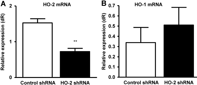
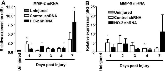
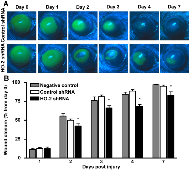
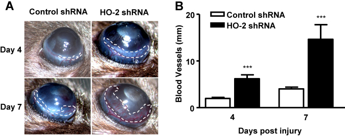
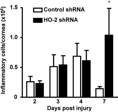
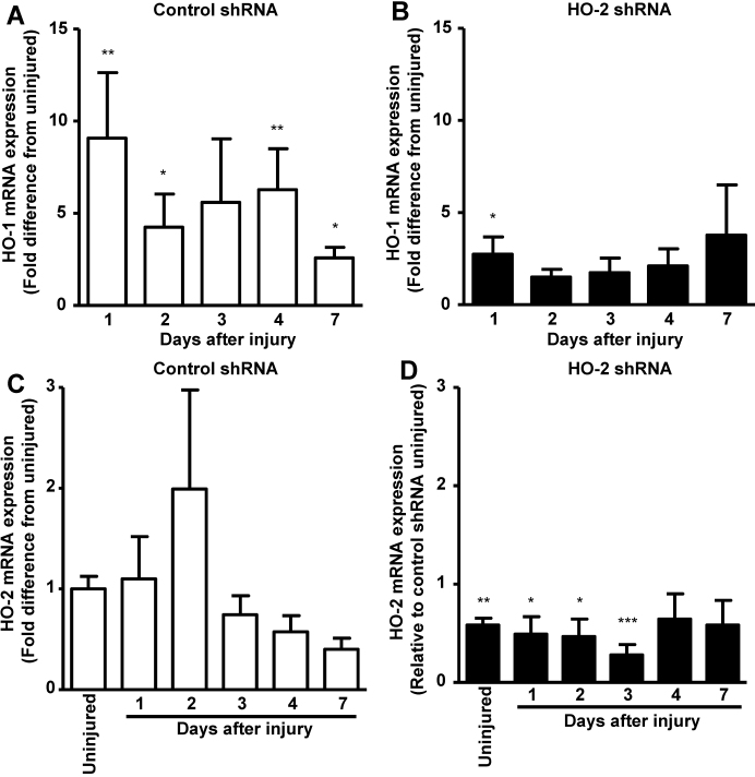
Local injection of HO-2-specific shRNA led to a 50% reduction in corneal HO-2 mRNA. Administration of HO-2-specific shRNA delayed corneal re-epithelialization when compared with the control shRNA-treated group by 14%, 20%, and 12% at days 3, 4, and 7 after injury, respectively (n=18-24). The observed delay in the wound repair process in HO-2 shRNA treated mice was accompanied by a threefold and 3.5 fold increase in the neovascular response at days 4 and 7 after injury. Further, local knockdown of HO-2 lead to an aberrant chronic inflammatory response, as shown by presence of high numbers of inflammatory cells still present in the cornea at day 7 after injury; 1.04±0.45×10(6) in HO-2 knockdown mice versus 0.14±0.03×10(6) inflammatory cells in control mice. Matrix metalloproteinase-2 (MMP-2) but not MMP-9 increased following injury and remained elevated in the injured corneas of the HO-2 shRNA-treated eyes.
Corneal knockdown of HO-2 via local administration of HO-2-specific shRNA leads to delayed re-epithelialization, increased neovascularization and an aberrant inflammatory response similar to what is observed in the HO-2 null mouse. The elevated MMP-2 expression may contribute to the increase in neovascularization in corneas in which HO-2 expression is suppressed.
血红素加氧酶(HO)-2在角膜上皮中高表达,是血红素加氧酶系统的一个组成部分,该系统基于其调节白细胞迁移以及通过其产物胆绿素/胆红素和一氧化碳(CO)抑制炎性细胞因子和蛋白质表达的能力,代表一种内在的细胞保护和抗炎系统。我们已经表明,在HO-2基因敲除小鼠中,上皮损伤会导致角膜炎症无法消退以及出现慢性炎症并发症,包括溃疡、穿孔和新生血管形成。在本研究中,我们探讨角膜局部HO-2抑制是否足以破坏角膜固有的抗炎和修复能力。
在角膜上皮清创术前一天开始,将针对HO-2的沉默发夹RNA(shRNA)结膜下注射(100 ng/眼)以及局部应用(100 ng/眼),此后每天一次。在麻醉的小鼠中使用Alger刷去除角膜上皮。使用解剖显微镜和图像分析通过荧光素染色评估再上皮化。通过髓过氧化物酶活性定量炎症反应。通过RT-PCR测量mRNA水平。
局部注射HO-2特异性shRNA导致角膜HO-2 mRNA减少50%。与对照shRNA处理组相比,给予HO-2特异性shRNA后,损伤后第3、4和7天角膜再上皮化分别延迟14%、20%和12%(n = 18 - 24)。在HO-2 shRNA处理的小鼠中观察到的伤口修复过程延迟伴随着损伤后第4天和第7天新生血管反应分别增加了3倍和3.5倍。此外,HO-2的局部敲低导致异常的慢性炎症反应,如损伤后第7天角膜中仍存在大量炎性细胞所示;HO-2敲低小鼠中为1.04±0.45×10⁶个炎性细胞,而对照小鼠中为0.14±0.03×10⁶个炎性细胞。基质金属蛋白酶-2(MMP-2)而非MMP-9在损伤后增加,并在HO-2 shRNA处理眼的损伤角膜中保持升高。
通过局部给予HO-2特异性shRNA敲低角膜中的HO-2会导致再上皮化延迟、新生血管形成增加以及出现与HO-2基因敲除小鼠中观察到的类似的异常炎症反应。MMP-2表达升高可能导致HO-2表达被抑制的角膜中新生血管形成增加。