Kwon Jin Sook, Park Rho Kwan, Shim Tae Jin, Jeong Myung Ho, Cho Myeong Chan, Ahn Youngkeun, Kim Dong-Woon
Department of Cardiology, Chonnam National University Hospital, Gwanju 501-757, Korea.
J Vet Sci. 2011 Jun;12(2):143-9. doi: 10.4142/jvs.2011.12.2.143.
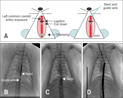
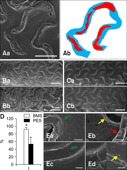
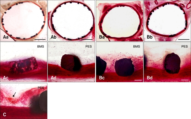
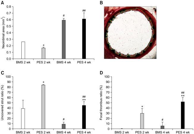
The purpose of our study was to create a novel rat aorta stent implantation model. Stainless steel bare metal stents (BMS) or paclitaxel-eluting stents (PES) were implanted in male Sprague-Dawley rats (BW 400 ± 20 g). Two and four weeks after stent implantation, the aorta were collected, fixed with 2% glutaraldehyde, and cut into two segments. One segment was used for scanning electron microscopy analysis to evaluate re-endothelialization, and the other segment was used to calculate the neointimal area. At 2 weeks after stenting, the appearance of neointimal hyperplasia was less in the PES group than in the BMS group. At 4 weeks after stenting, no significant difference in neointimal hyperplasia was observed between two groups. On the other hand, the PES group showed more thrombus formation and less re-endothelialization compared to the BMS group. This study demonstrated the ability of a novel rat model of aorta stenting via a common carotid artery to measure the efficacy and safety of commercially available drug-eluting stents.
我们研究的目的是创建一种新型大鼠主动脉支架植入模型。将不锈钢裸金属支架(BMS)或紫杉醇洗脱支架(PES)植入雄性Sprague-Dawley大鼠(体重400±20 g)体内。支架植入后2周和4周,采集主动脉,用2%戊二醛固定,切成两段。一段用于扫描电子显微镜分析以评估再内皮化,另一段用于计算新生内膜面积。支架植入后2周,PES组新生内膜增生的表现比BMS组少。支架植入后4周,两组之间新生内膜增生未观察到显著差异。另一方面,与BMS组相比,PES组显示出更多的血栓形成和更少的再内皮化。本研究证明了一种通过颈总动脉进行主动脉支架置入的新型大鼠模型能够测量市售药物洗脱支架的疗效和安全性。